Download
1 / 18
190 likes | 441 Views
1. Free energy with controlled uncertainty 2. The modes of ligand binding to DNA. Tomáš Kubař Institute of Organic Chemistry and Biochemistry Praha, Czech Republic. Thermodynamic Integration. ‘Alchemical’ change of an atom/group Force-field parameters for both initial and final state

E N D
1. Free energy with controlled uncertainty2. The modes of ligand binding to DNA Tomáš Kubař Institute of Organic Chemistry and Biochemistry Praha, Czech Republic
Thermodynamic Integration • ‘Alchemical’ change of an atom/group • Force-field parameters for both initial and final state • Coupling parameter l introduced – mixing of the initial (l=0) and final (l=1) states: • Simulation for a number of l values from 0 to 1; then, for every l: • Finally, numerical integration gives the free-energy difference of the initial and final state:
Usual simulation protocol • In every l point (window) – a simulation with separated equilibration period and production phase, both of fixed length • Only the production phase used to calculate dG/dl • The uncertainty of dG/dl may be evaluated but not controlled in every lpoint dV/dl values discarded calculated mean value of dV/dl
Reverse cumulative averaging • No preset length of equilibration and production phase • We set the requested maximum uncertainty of dG/dl and DG • Important – determine the equilibrated phase of simulation • Criterion for thermodynamic equilibrium: dV/dl values come from a normal distribution • The production region determined as the longest equilibrated tail of the simulation – therefore “reverse” • Standard error of the mean <dV/dl> is calculated in the production region • IF error > threshold THEN continue with current l ELSE record dG/dl = <dV/dl> and proceed to the next l dV/dl values discarded calculated mean value of dV/dl and its error JCP 2004, 120, 2618
Reverse cumulative averaging • The simulation proceeds in blocks of fixed length (2000 steps) • RCA takes place after a block finishes • Shapiro–Wilk normality test adopted from the R project (GPL) • Normality determined on the 85% confidence level • A fragment of the .mdp file: free-energy-method = ti_rca init_lambda = 0.0 delta_lambda = 0.05 lambda_points = 21 target_error = 5.0 – requested maximum uncertainty of dG/dl(kJ/mol) – thus, the upper bound of uncertainty of total DG
Application IDNA intercalation • Ethidium – DNA binding drug, a strong carcinogen • Binding free energy difference of ethidium and its derivatives CEJ 2006, 12, 280
Application IDNA intercalation • Thermodynamic cycle DNA…ETD Free ETD DNA…EPP Free EPP DDG = DG(2) – DG(1) = DG(B) – DG(A) • Results: exptl: +1.6 kcal/mol 1 A B 2
Application IIThermostable protein and its mutants • Rubredoxine – a globular protein, which survives temperatures over 100 °C – containts a distinct hydrophobic core – mutation of a bulky amino acid in the core makes melting temperature drop • Folding free energy difference of RB and the mutants • Results may be compared with a calorimetric experiment CEJ 2007, submitted
Application IIThermostable protein and its mutants • Thermodynamic cycle Denat-WT Folded-WT Denat-F48A Folded-F48A DDG = DG(2) – DG(1) = DG(B) – DG(A) • Studied mutations: 1 A B 2 F48A F48G WT (F29A) F29G F29I
Modes of ligand binding to DNA • The most important non-covalent binding modes: intercalation minor-groove binding • What do they have in common? • What are the distinctions between them? (apart from the obvious – the deformation of DNA)
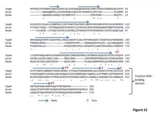
The molecular systems and force field • DNA – two decanucleotides • (CGTATATACG)2 – AT rich • (CGCGCGCGCG)2 – CG rich • The ligand – ellipticin and its derivatives (9-hydroxy – polar, and also both forms protonated on N2) • Force field – AMBER parm99 + parmBSC0 + TIP3P H2O • For every complex – a 50ns simulation at 300 K, 1 atm
Look at the trajectory • Intercalative complexes • all stable, the ligand remains in the binding site • Minor-groove complexes • all molecules with AT-rich – stable • all molecules with GC-rich – unstable • the ligand leaves the minor groove; either flows away from the DNA or gets stacked at the end of the double helix • First conclusions – sequence preference • intercalative mode – none or weak • minor-groove binding – strong preference for AT-rich • We should look for an explanation… later on
The interaction energy • The measure of inherent attraction between molecules, without any external influence (environment) • energygrps in .mdp • protonated × neutral molecules • otherwise, little significant difference found
Quantification of the dynamics of complex • The desired quantity is entropy (as component of free energy) • How to calculate entropy: trajectory –> covariance matrix of atomic fluctuations –> eigenvalues –> normal-mode vibration frequency –> entropy (following Schlitter CPL 215, 617 (1993) or Karplus JCP 115, 6289 (2001)) • Using g_covar • Entropy depends on the length of simulation upon which the covariance matrix was constructed – convergence behavior should be checked • Only the heavy atoms of DNA and ligand involved
Conformational entropy • intercalative complexes favored by –TDS≈ 30 kcal/mol • which part of the complex experiences different dynamics?
Dynamics of the complex • A look at the dynamic behavior of individual nucleotides: g_rmsf • The change in flexibility of DNA is localized in a few nucleotide residues
Why no MiG binding to GC-rich? • The reason must be sought in the DNA itself, not the ligand • MiG width is the same in AT-rich and GC-rich (not shown) • Interaction energy of bases with water in bare DNA…? kcal/mol AT-rich –44 GC-rich –63 • This may be the explanation. The (any?) ligand cannot probably compete with water in the binding to DNA bases.