Download
1 / 56
1.18k likes | 6.22k Views
CHEST WALL DEFORMITIES. Carlos A. Angel, MD. CHEST WALL DEFORMITIES. Pectus excavatum Pectus carinatum Poland syndrome Sternal defects Rare lesions: Thoracic ectopia cordis Jeune asphyxiating thoracic dystrophy . PECTUS EXCAVATUM.

E N D
CHEST WALL DEFORMITIES Carlos A. Angel, MD
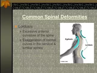
CHEST WALL DEFORMITIES • Pectusexcavatum • Pectuscarinatum • Poland syndrome • Sternal defects • Rare lesions: Thoracic ectopiacordis Jeune asphyxiating thoracic dystrophy
PECTUS EXCAVATUM • Most common anterior chest wall deformity (7-38/10,000 births) • Positive family history (37%-47%) • 3:1 M:F ratio • Spontaneous resolution is rare • Progression is expected during growth spurts • Tall, lanky , poor posture • Cause unknown • Can be acquired after correction of CDH.
PRESENTATION • Clinical spectrum • Posterior angulation of the body of the sternum • Posterior angulation of the costal cartilages that meet the sternum • In severe cases posterior angulation of the most anterior portion of the osseous ribs • Depression may be assymetric (carinatum/excavatum deformities) • Currarino- Silvermann deformity ( protrusion of sterno-manubrial joint)
PRESENTATION • Many are asymptomatic • Precordial pain • Pain after sustained exercise • Palpitation (mitral valve prolapse) • Systolic ejection murmur is frequently identified • Shortness of breath • Decreased exercise tolerance
Associated Abnormalities • 704 patients Scoliosis 107 kyphosis 4 Myopathy 3 Poland syndrome 3 Marfan syndrome 2 Pierre Robin syndrome 2 Prune belly syndrome 2 Neurofibromatosis 3 Cerebral palsy 4 Tuberous sclerosis 1 CDH 1 Shamberger RC, Welch KJ,: Surgical repair of pectusexcavatum. J PediatrSurg 1998; 23:615-622
PECTUS EXCAVATUM • Some believe this is a purely cosmetic condition • This contrasts with the clinical impression that many patients report improved breathing, stamina and exercise tolerance after repair • Despite 6 decades of work, no consensus has been achieved as to what degree of cardiopulmonary impairment is present, if any, in patients with depression chest wall deformities
PECTUS EXCAVATUM • Work-up • CT of the chest • Pulmonary Function Tests • Echocardiogram • Type and crossmatch PRBC’s
PULMONARY FUNCTION Castile et. al., ( 8 pts, 1 carinatum) • MeanTLC 79% of predicted • No suggestion of a significant ventilation-perfusion abnormality • With maximum workload oxygen extraction exceeded predicted values in symptomatic patients • Increases in tidal volume with exercise were uniformly depressed • No postoperative studies performed
PULMONARY FUNCTION • Brown et.al. Respiratory studies before and after surgery Vital Capacity- nl Maximum breathing capacity greater than 50%decreased (9/11 pts), increased 31% after repair • Orzaleski and Cook 12 children with severe pectusexcavatum deformities Significant decrease (p <0.001) in VC, TLC and maximal breathing capacity • Lise and Buhlmann Pre and pop lung volumes in 12 adults (3-11 y after operation) Absolute lung volume only improved in patients with interval increase in height Work capacity increased in 9/10 patients Pop decrease in heart rate at a given power output Some of the improvement may have resulted from increased cardiac stroke volume
PULMONARY FUNCTION • Cahill et.al. 19 children and adolescents (5 carinatum, 14 excavatum) No pre-0p or pop abnormalities seen in carinatum patients Excavatum patients showed low normal VC, unchanged by operation Operation changed TLC Significant improvement after operation in in maximum voluntary ventilation and exercise tolerance • Devereaux et.al. 88 pts with pectusexcavatum 1-20yrs after operation (avg 8 yrs) Those with <75% predicted function pre-operatively had improved function Those with >75% had worsening function, this was in contrast with subjective reports of improvement in symptoms
PULMONARY FUNCTION • Wynn et.al. 12 children Decline in TLC after repair • Kaguraoka et.al. 138 pts Temporary decrease in pulmonary function after surgery • Haller et.al. 36 pts pectusexcavatum, 10 controls Decreased FVC did not change after repair Improved exercise tolerance after repair in 66% of patients, likely the result of improved cardiac function
PULMONARY FUNCTION Minimally Invasive Repair Studies: • Borowitz et.al. 10 pts Normal pulmonary function pre and pop • Sigalet et. al. 11 pts Subjective improvement in exercise tolerance Pulmonary function significantly reduced at 3mo. Cardiac function enhanced with increase stroke volume Limitation in exercise had a cardiovascular rather than a pulmonary cause • Lawson et.al. 408 pectusexcavatum patients 45 PFT’s after Nuss procedure and bar removal Pre-operative values for FVC, FEV1 and forced expiratory flow were 13-20% below average Post-operative significant improvement for al parameters greatest gains by surgery were seen in patients older than 11 yrs
PULMONARY FUNCTION CONCLUSIONS: In the last decade , studies of hundreds of patients with pectusexcavatum have demonstrated that it is associated with an average decrease of pulmonary function of 85% of predicted values ( 80% is 2 SD below the norm). The increase in function after surgery occurs in patients with normal pulmonary parenchyma and airways
CARDIAC FUNCTION • Deformity of the heart • Sternal imprint of the anterior R ventricle • Displacement of the heart to the L side • Garusi, et.al. Decreased work capacity significantly lower in sitting than in supine position Stroke volume decreased 40.3% from supine to sitting position Increased cardiac output is achieved by increased heart rate, not stroke volume • Beiser et. al.- Provided further evidence that cardiac function is impaired during upright exercise
CARDIAC FUNCTION • Peterson et.al. 13 patients with pectusexcavatum (11 symptomatic) Radionuclide angiography Marked decrease in symptoms during exercise after surgical correction during a regulated exercise protocol No changes in L ventricular EF • Kowaleski et. al 42 pts Echocardiographic evaluation of cardiac function Statistically significant changes seen in RV indices (systolic, diastolic and stroke volume) after surgery All limitations in stroke volume result from R ventricular compression
CARDIAC FUNCTION • Echocardiographic studies: Mitral valve prolapse 18% ( Udoshi et.al., CHKD, Norfolk) 65% ( Saint- Mezard et.al.) • Resolution of prolapse after repair seen in 43-44%
BODY IMAGE • Large percentage of patients are self-concious about their chests • Even suicide attempts have been reported • Not an inconsequential problem • Psychometric assessments in more than 300 children- Marked improvement in psychosocial functioning after repair • Severity of deformity did not correlate with the parents/patients perception of body image concerns • Pectusexcavatum is a deformity which worsens during a developmental period in which body image is crucial
INDICATIONS • Progressive symptoms • Restrictive disease, decreased work production or oxygen uptake as demonstrated by PFT’s • Ct scan showing cardiac compression or displacement • Haller index greater than 3.25 • Pulmonary atelectases • Mitral valve prolapse, bundle branch block • Recurrent pectusexcavatum after repair
TIMING • Can be performed in younger children with severe exercise tolerance • Best deferred until after the pubertal growth spur
RAVITCH PROCEDURE • Transverse skin incision • Mobilization and retraction of pectoralis and rectus abdominis muscles • Excision of deformed cartilagues leaving the perichondrium intact • Fracture of the sternum (wedge osteotomy) • Metal strut for stabilization
COMPLICATIONS Early: • Wound infection (1%) • Pneumothorax (4%) • Hemothorax (0.6%) • Pneumonia (0.5%) • Pericarditis (0.4%) • Pleural effussion (0.3%) Late: • Bar infection (0.5%, only 0.2% required removal) • Bar displacement (1% -5.7%) • Nickel allergy (3%) • Recurrence • Repairs performed in children <4yo result in impaired growth of the ribs resulting in a band-like narrowing of the chest the chest
POST-OPERATIVE PERIOD • 5-7 days in the hospital • Peri-operative antibiotics • Pain management ( epidural PCA, IV acetaminophen, ketoralac) • DVT prophylaxis • Incentive spirometer • Muscle relaxants (diazepam, methocarbamol) • H2 blockers
BAR REMOVAL • Bar stays in place a minimum of 2 years • Bar should be left in longer in younger patients • Patients evaluated on an annual basis • Exercise program very important • Removal is an outpatient surgery procedure under GA
NUSS vs. RAVITCH • Meta-analysis (JPS May 2010) • No prospective randomized trials • 9 prospective and retrospective studies • No significant difference in complication rates • Rate of reoperation because of bar migration or persistent deformity higher in Nuss • POP pneumothorax or hemothorax higher in Nuss • Duration of operation longer in Ravitch • No difference in length of hospital stay, time to ambulation, or patient satisfaction.
POST-OPERATIVE ACTIVITY • Deep- breathing 2x day • Bathe or shower after 5 days • No waist bending, twisting or log rolling for the first 4 weeks • No slouching first month • No heavy lifting for 2 months • No contact sports for 3 mo Other recommendations: • MedAlert bracelet recommenced • May have MRI • Cardiac defibrillation with anterior posterir paddle placement
BAR REMOVAL • Usually performed after 2 years • General anesthesia • Day surgery procedure • Chest Xray recommended
PECTUS CARINATUM • Less frequent than pectusexcavatum • M:F 4:1 • No known cause • Mild deformity at birth worsens as the child grows • Positive family history in 26% • History of scoliosis in 15%
PECTUS CARINATUM Presentation: • Symmetric or asymetric protrusion of the sternum • Associated lateral depression of the ribs • Pain in the area • Some patients experience exercise limitation • Rotation of the sternum is often seen • Chondromanubrial protrusion (rare), results in a comma-shaped sternum ( these children have andincreased incidence of heart disease)