Download
1 / 83
850 likes | 1.12k Views
Today's Medical Assistant 2 th edition. Chapter 07 Skeletal System. Overview of the Skeletal System. List and describe five functions of the skeletal system. Explain the difference between compact and spongy bone. Classify bones according to size and shape. Lesson 7.1.

E N D
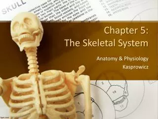
Today's Medical Assistant 2th edition Chapter 07 Skeletal System
Overview of the Skeletal System List and describe five functions of the skeletal system. Explain the difference between compact and spongy bone. Classify bones according to size and shape. Lesson 7.1
Identify the general features of a long bone. Explain the process by which long bones grow in length. Explain the difference between the axial and appendicular skeletons. Lesson 7.1 Overview of the Skeletal System (cont’d)
Introduction to the Skeletal System Consists of: Bones and cartilage Ligaments Tendons associated with bones
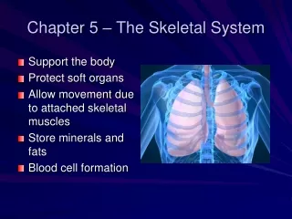
Functions of the Skeletal System Support Provides a rigid framework Supports the soft organs of the body Protection Protects the soft body parts Cranium protects the brain Vertebrae protect the spinal cord Rib cage protects heart and lungs
Functions of the Skeletal System • Movement • Bones and muscles work together to produce body movement • Storage • Calcium: needed for vital metabolic processes • When blood calcium levels decrease: calcium is released from the bones • When blood calcium levels increase: excess calcium is stored in the bones • Fat is stored in the yellow bone marrow
Functions of the Skeletal System • Blood cell formation • Hematopoiesis: blood cell formation (red blood cells, white blood cells, platelets) • Takes place mostly in the red bone marrow • Found in most bones in an infant • With age: largely replaced by yellow marrow (fat) storage
Structure of Bone Tissue • Compact bone • Osteon (haversian system): the microscopic unit of compact bone • Packed tightly together to form a solid mass • Osteonic (haversian) canal: a central canal in the osteon • Contains a blood vessel
Structure of Bone Tissue • Compact bone • Lamellae: concentric rings of hard calcified matrix that surrounds osteonic canals • Osteocytes: bone cells • Lacunae: spaces between the rings of matrix, which contain bone cells (osteocytes) • Canaliculi: small channels that radiate from the lacunae to the osteonic canal • Provide passageways through the hard matrix
Structure of Bone Tissue • Spongy (cancellous) bone • Lighter and less dense than compact bone • Consists of plates of bone (trabeculae)
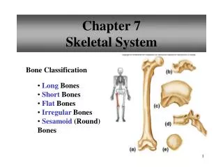
Classification of Bones • Long bones: longer than they are wide • Consist of a long shaft with two bulky ends • Primarily compact bone • May have a large amount of spongy bone at the ends • Examples: thigh, leg, arm, and forearm • Short bones: cube shaped • Consist primarily of spongy bone • Covered by a thin layer of compact bone • Examples: bones of wrist and ankle
Classification of Bones • Flat bones: thin, flattened, and often curved • Arranged similar to a sandwich • Middle layer of spongy bone covered on each side by a layer of compact bone • Example: most of the bones of the cranium • Irregular bones • Primarily spongy, covered with a thin layer of compact bone • Examples: vertebrae and some skull bones
General Features of a Long Bone • Diaphysis: shaft of a long bone • Consists of compact bone • Medullarycavity: space inside the shaft of a long bone • In adults: contains yellow bone marrow
General Features of a Long Bone • Epiphysis: the expanded portion at the end of a long bone • Spongy bone covered by a thin layer of compact bone • Articular cartilage: thin layer of hyaline cartilage that covers the ends of long bones • Provides smooth surfaces for movement in the joints
General Features of a Long Bone • Epiphyseal plate: plate of hyaline cartilage between the diaphysis and epiphysis • Bones grow in length at the epiphyseal plate • Growth ceases when the cartilaginous epiphyseal plate is replaced by a bony epiphyseal line • Periosteum: tough, fibrous connective tissue that covers a long bone except in the region of the articular cartilage • Richly supplied with nerve fibers, lymphatic vessels, blood vessels, and osteoblasts
General Features of a Long Bone • Nutrient foramina: small openings in the diaphysis of the bone for the passage of blood vessels • Endosteum: thin connective tissue membrane that lines the medullary cavity
Bone Development and Growth • Osteogenesis (also known as ossification): process of bone formation • Cells involved: • Osteoblasts: bone-forming cells • Osteocytes: mature bone cells • Osteoclasts: break down and reabsorb bone
Bone Development and Growth • Bone growth in length • Hyaline cartilage in epiphyseal plate: grows by mitosis • Chondrocytes next to diaphysis age and degenerate • Osteoblasts ossify the matrix to form bone • Continues throughout childhood and adolescence • Cartilage growth ceases: usually in early 20s • Epiphyseal plate completely ossifies • Epiphyseal line remains • Bones can no longer grow in length
Bone Development and Growth • Bone growth in length • Bone growth under influence of: • Growth hormone (secreted by anterior pituitary) • Sex hormones (secreted by ovaries and testes
Division of the Skeleton • Adult skeleton: consists of 206 bones • Axial skeleton: 80 bones, which include bones of head, vertebral column, ribs, sternum • Appendicular skeleton: 126 bones, which include the free appendages and their attachments to the axial skeleton
Bones and Articulations Identify the bones of the skull. Identify the structural features of vertebrae. List and describe the divisions of the vertebral column. Describe the structural features of the sternum and ribs. Lesson 7.2
Identify the parts of the pectoral girdle. Identify the bones of the upper extremities. Identify the parts of the pelvic girdle. Identify the bones of the lower extremities. Lesson 7.2 Bones and Articulations (cont’d)
List and describe the different types of joints. Describe ways in which the aging of an individual affects the skeletal system. Identify pathology related to the skeletal system. Lesson 7.2 Bones and Articulations (cont’d)
Skull • Skull • Made up of 28 bones • Cranium • Houses the brain
Cranium • Frontal bone • Paranasalfrontal sinuses: air-filled cavities in the frontal bone • Reduce the weight of the skull • Parietal bones • Joined to each other in the midline by the sagittal suture • Joined to the frontal bone by the coronal suture
Cranium • Occipital bone • Joined to the parietal bones by the lambdoidsuture • Foramen magnum: large opening on the lower surface of the occipital bone • Spinal cord passes through this opening • Occipital condyles: rounded processes on each side of the foramen magnum • Articulate with the first cervical vertebra
Cranium • Temporal bones • Meet the parietal bone at the squamous suture • External auditory meatus: canal that leads to the middle ear • Mandibularfossa: articulates with the mandible • Mastoid process: contains air cells that drain into middle ear cavity • Zygomatic process: helps form the prominence of the cheek
Cranium • Sphenoid bone • Spans the entire width of the cranial floor • Optic foramina: two openings for the passage of the optic nerve • Contains paranasalsphenoid sinuses • Ethmoid bone • Forms most of the bony area between the nasal cavity and the orbits • Contains paranasalethmoidal sinuses
Facial Bones • Form the basic framework and shape of the face • Maxillary bones • Forms upper jaw • Alveolar process: tooth socket • Palatine bones • Form posterior portion of hard palate and lateral walls of nasal cavity • Nasal bones • Form the bridge of the nose
Facial Bones • Lacrimal bones • Lacrimal groove: pathway for a tube that carries tears from eyes to nasal cavity • Zygomatic bones • Form the prominences of the cheeks • Temporal process that forms the zygomatic arch • Inferior nasal conchae • Thin, curved bones attached to the lateral walls of the nasal cavity
Facial Bones • Vomer • Helps to form the nasal septum • Mandible • Forms the lower jaw • Mandibularcondyle fits into mandibularfossa of the temporal bone to form the temporomandibular joint • Alveolar process: tooth socket
Auditory Ossicles • Malleus, incus, stapes • Transmit sound waves from the tympanic membrane to inner ear
Hyoid Bone • U-shaped bone in the neck • Located between the mandible and the larynx
Hyoid Bone • Only bone in the body that does not articulate directly with another bone • Functions: • Serves as a base for the tongue • Attachment for several muscles associated with swallowing
Vertebral Column • Extends from skull to pelvis • Contains 26 vertebrae • Intervertebral discs: pads of fibrocartilage that separate vertebrae • Function • Shock absorbers • Allow the vertebral column to bend
Vertebral Column • Four curvatures: increase strength and resilience of the column • Cervical • Thoracic • Lumbar • Sacral
General Structure of Vertebrae • Body (centrum): weight-bearing portion • Vertebral arch: posterior curved portion is the vertebral arch • Vertebral foramen: central large opening • When all the vertebrae are stacked together in a column • Vertebral foramina make a canal that contains the spinal cord
General Structure of Vertebrae • Transverse processes: project laterally from the vertebral arch • Place for muscle attachment • Spinous process: projects from the posterior midline • Place for muscle attachment • Can be felt as bony projections along the midline of the back
Composition of the Vertebral Column • Cervical vertebrae: C1-C7 • Thoracic vertebrae: T1-T12 • Lumbar vertebrae: L1-L5 • Make up the small of the back • Consist of large, heavy bodies • Support most of body weight • Have many back muscles attached to them
Composition of the Vertebral Column • Sacrum: triangular bone just below the lumbar vertebrae • Forms posterior wall of pelvic cavity • Coccyx (tailbone): last part of vertebral column
Thoracic Cage • Protects heart, lungs, and great vessels • Supports bones of the shoulder girdle • Plays a role in breathing • Components: • Thoracic vertebrae dorsally • Ribs laterally • Sternum and costal cartilage anteriorly
Sternum • Consists of three parts: • Manubrium: superior • Jugular (suprasternal) notch: indentation in the superior margin of the manubrium • Articulates with clavicles and first two pairs of ribs • Body: middle • Sternal angle: where manubrium and body meet (felt as a horizontal ridge) • Xiphoid process: inferior
Ribs • Twelve pairs of ribs • One pair attached to each of 12 thoracic vertebrae • True ribs: upper seven pairs • Attach to sternum directly by costal cartilage