Download
1 / 42
420 likes | 703 Views
PART 1. Basic Embryology. Embryology. Embryology – study of the origin and development of single individual Prenatal period Embryonic period – first 8 weeks Fetal period – remaining 30 weeks. Embryonic Period. Figure 3.1 (1 of 2). Fetal Period. Figure 3.1 (2 of 2).

E N D
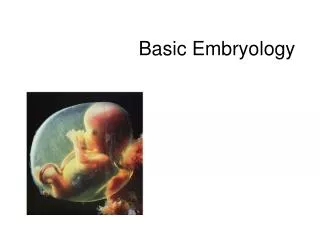
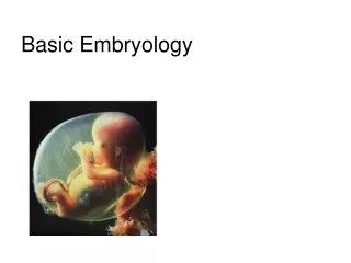
PART 1 Basic Embryology
Embryology • Embryology – study of the origin and development of single individual • Prenatal period • Embryonic period – first 8 weeks • Fetal period – remaining 30 weeks
Embryonic Period Figure 3.1 (1 of 2)
Fetal Period Figure 3.1 (2 of 2)
The Basic Body Plan • Skin – dermis and epidermis • Outer body wall – trunk muscles, ribs, vertebrae • Body cavity and digestive tube (inner tube) • Kidneys and gonads – deep to body wall • Limbs
The Basic Body Plan Figure 3.2
The Embryonic Period • Week 1 – from zygote to blastocyst • Conception – in lateral third of uterine tube • Zygote (fertilized oocyte) moves toward the uterus • Blastomeres – daughter cells formed from zygote • Morula – solid cluster of 12–16 blastomeres • “Mulberry” • Blastocyst – fluid-filled structure – ~ 60 cells
The Embryonic Period • Stages of first week • Zygote • 4-cell • Morula • Early blastocyst • Late blastocyst (implants at this stage)
Fertilization and the Events of the First 6 Days of Development Figure 3.3
Week 2 – The Two-Layered Embryo • Bilaminar embryonic disc – inner cell mass divided into two sheets • Epiblast and the hypoblast • Together they make up the bilaminar embryonic disc
Week 2 – The Two-Layered Embryo • Amniotic sac – formed by an extension of epiblast • Outer membrane forms the amnion • Inner membrane forms the amniotic sac cavity • Filled with amniotic fluid
Week 2 – The Two-Layered Embryo • Yolk sac – formed by an extension of hypoblast • Digestive tube forms from yolk sac • NOT a major source of nutrients for embryo • Tissues around yolk sac • Gives rise to earliest blood cells and blood vessels
Implantation of the Blastocyst Figure 3.4 (1 of 3)
Implantation of the Blastocyst Figure 3.4 (2 of 3)
Implantation of the Blastocyst Figure 3.4 (3 of 3)
Week 3 – The Three-Layered Embryo • Primitive streak – raised groove on the dorsal surface of the epiblast • Gastrulation – a process of invagination of epiblast cells • Begins at the primitive streak • Forms the three primary germ layers
Week 3 – The Three-Layered Embryo • Three Germ Layers* • Endoderm – formed from migrating cells that replace the hypoblast • Mesoderm – formed between epiblast and endoderm • Ectoderm – formed from epiblast cells that stay on dorsal surface *All layers derive from epiblast cells!
The Primitive Streak Figure 3.5e–h
The Notochord • Primitive node – a swelling at one end of primitive streak • Notochord forms from primitive node and endoderm • Notochord – defines body axis • Is the site of the future vertebral column • Appears on day 16
Formation of the Mesoderm and Notochord Figure 3.6
Neurulation • Neurulation – ectoderm starts forming brain and spinal cord • Neural plate – ectoderm in the dorsal midline thickens • Neural groove – ectoderm folds inward
Neurulation • Neurulation (continued) • Neural tube – a hollow tube pinches off into the body • Cranial part of the neural tube becomes the brain • Maternal folic acid deficiency causes neural tube defects
Neurulation • Neural crest • Cells originate from ectodermal cells • Forms sensory nerve cells • Induction • Ability of one group of cells to influence developmental direction of other cells
The Mesoderm Begins to Differentiate • Somites – our first body segments • Paraxial mesoderm • Intermediate mesoderm – begins as a continuous strip of tissue just lateral to the paraxial mesoderm
The Mesoderm Begins to Differentiate • Lateral plate – most lateral part of the mesoderm • Coelom – becomes serous body cavities • Somatic mesoderm – apposed to the ectoderm • Splanchnic mesoderm – apposed to the endoderm
PART 2 Basic Embryology
Changes in the Embryo Figure 3.7a, b
Changes in the Embryo Figure 3.7c, d
Week 4 – The Body Takes Shape • Folding of embryo laterally and at the head and tail • Embryonic disc bulges; growing faster than yolk sac • “Tadpole shape” by day 24 after conception • Primitive gut – encloses tubular part of the yolk sac • Site of future digestive tube and respiratory structures
Week 4 – The Body Takes Shape Figure 3.8
Week 4 – The Body Takes Shape • Derivatives of the germ layers • Ectoderm forms • Brain, spinal cord, and epidermis • Endoderm forms • Inner epithelial lining of the gut tube • Respiratory tubes, digestive organs, and urinary bladder • Notochord – gives rise to nucleus pulposus within intervertebral discs
Week 4 – The Body Takes Shape • Mesoderm – forms • Muscle • Bone • Dermis • Connective tissues (all) • Mesoderm differentiates further and is more complex than the other two layers
Derivatives of Germ Layers Figure 3.10
Week 4 – The Body Takes Shape • Mesoderm (continued) • Somites divides into • Sclerotome • Dermatome • Myotome • Intermediate mesoderm forms • Kidneys and gonads
Week 4 – The Body Takes Shape • Mesoderm (continued) • Splanchnic mesoderm • Forms musculature, connective tissues, and serosa of the digestive and respiratory structures • Forms heart and most blood vessels • Somatic mesoderm – forms • Dermis of skin • Bones • Ligaments
PART 3 Basic Embryology
The Germ Layers in Week Four Figure 3.9a–d
Week 5-8 – The Second Month of Embryonic Development • Limb buds form • Embryo first looks recognizably human (week 8) • Head is disproportionately large • All major organs are in place Figure 3.11
The Fetal Period • A time of maturation and rapid growth • Cells are differentiating during the first half of the fetal period • Normal births occur 38 weeks after conception • Premature birth is one that occurs before 38 weeks PLAY Ultrasound of Fetus
Developmental Events of the Fetal Period Table 3.1 (1 of 3)
Developmental Events of the Fetal Period Table 3.1 (2 of 3)
Developmental Events of the Fetal Period Table 3.1 (3 of 3)