Download
1 / 60
630 likes | 1.09k Views
Analysis of Gene Expression Data. Rainer Breitling r.breitling@bio.gla.ac.uk Bioinformatics Research Centre and Institute of Biomedical and Life Sciences University of Glasgow. Outline. Gene expression biology Measuring gene expression levels

E N D
Analysis of Gene Expression Data Rainer Breitling r.breitling@bio.gla.ac.uk Bioinformatics Research Centre and Institute of Biomedical and Life Sciences University of Glasgow
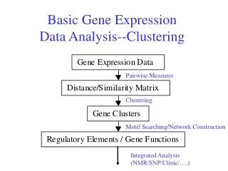
Outline • Gene expression biology • Measuring gene expression levels • two technologies: Two-color cDNA arrays and single-color Affymetrix genechips • Finding and understanding differentially expressed genes • Advanced analysis (clustering and classification) • Cutting-edge uses of microarray technology
...but the complexity and diversity of the resulting phenotype is challenging whole-mount in situ hybridization of X. laevis tadpoles
The dramatic consequences of gene regulation in biology • Same genome • Different tissues • Different physiology • Different proteome • Different expression pattern Anise swallowtail, Papilio zelicaon
Regulatory Networks – integrating it all together Genetic regulatory network controlling the development of the body plan of the sea urchin embryo Davidson et al., Science, 295(5560):1669-1678.
Gene expression distinguishes... • ...physiological status (nutrition, environment) • ...sex and age • ...various tissues and cell types • ...response to stimuli (drugs, signals, toxins) • ...health and disease • underlying pathogenic diversity • progression and response to treatment • patient classes of varying prospects
Measuring gene expression levels • total amount of mRNA = optical density at appropriate (UV) wavelength • mass separation and specific probing, one gene at a time = Northern blot • comprehensive “molecular sorting” = microarray technology • two-color cDNA or oligo arrays • single-color Affymetrix genechips
cDNA microarray schema color code for relative expression From Duggan et al.Nature Genetics21, 10 – 14 (1999)
cDNA microarray raw data • can be custom-made in the laboratory • always compares two samples • relatively cheap • up to about 20,000 mRNAs measured per array • probes about 50 to a few hundred nucleotides Yeast genome microarray. The actual size of the microarray is 18 mm by 18 mm. (DeRisi, Iyer & Brown, Science, 268: 680-687, 1997)
GeneChip® Hybridization Image courtesy of Affymetrix.
Affymetrix genearrays single color (color code indicates only hybridization intensity) high density, perfectly addressable probes multiple probes per gene/mRNA
Affymetrix genechips contain “probe sets” instead of single probes per gene better reliability of the results (each probe is [almost] an independent test)
Mismatch probes allow present/absence calls for every single probe set PM probes MM probes Wilcoxon Signed Rank Test : non-parametric test; Take the paired observations (PM-MM), calculate the differences, and rank them from smallest to largest by absolute value. Add all the ranks associated with positive differences, giving the T+ statistic. Finally, the p-value associated with this statistic is found from an appropriate table. (MathWorld)
Scatter plots classical scatter plot M-A plot for microarray analysis M A Differentially expressed genes are higher (or lower) in one of the samples Use an appropriate cut-off (‘distance’ from diagonal) to select relevant genes highly arbitrary!
t-test = statistical significance of observed difference • requires independent experimental replication • assumes the data are identically normally distributed
Sample 2 Sample 1 Frequency Probability 0 1 2 3 -3 -2 -1 Testing an intrinsic hypothesis • Two samples (1, 2) with mean expression that differ by some amount d. • If H0:d= 0 is true, then the expected distribution of the test statistic t is
Volcano plot Scatter plot of -log(p-value) from a t-test vs. log ratio. Visualises fold-change and statistical significance at the same time: Find genes that are significant and have large fold change, and genes that are significant but have small fold change.
Is this gene changed? Comparison with all other genes on the array Expression of gene A • Rank Product: • RP = (3/10) * (1/10) * (2/10) * (5/10) • intuitive • non-parametric, powerful test statistic • more reliable detection of changed genes in noisy data with few replicates Significance estimate based on random permutations: Probability that gene A shows such an effect by chance: p ≤ 0.03 Expectation to see any gene (out of 10) with such a effect: E-value ≈ 0.5 Breitling et al., FEBS Letters, 2004
Multiple Testing Problem • microarrays measure expression of >10,000 genes at the same time many thousands of statistical tests are performed • type 1-error: Calling a gene significantly changed, even if it’s just by chance protect yourself by Bonferroni correction • type 2-error: Missing a significantly changed gene reduce this problem by Benjamini-Hochberg false-discovery rate procedure
Multiple Testing Problem Bonferroni correction. n independent tests, control the probability that a spurious result passes the test at signficance level α adjust acceptance level for each individual test as: Benjamini-Hochberg False Discovery Rate. Control the number of false positives (N1|0) among the top R genes at the significance level α.
The result of “differential expression” statistical analysis a long list of genes!
Biological Interpretation Strategy • Are certain types of genes more common at the top of the list and is that significant? • Challenges: • Some types of genes are more common in the genome/on the array • The list of genes usually stops at an arbitrary cut-off (“significantly changed genes”) • Classifying genes according to “gene type” is a tedious task • Expectations and focused expertise might bias the interpretation • Early discoveries might restrict further analysis • Solution: Automated procedure using available annotations
iterative Group Analysis (iGA) iGA uses a simple hypergeometric distribution to obtain p-values Breitling et al. (2004), BMC Bioinformatics, 5:34.
Possible sources of classification • adjacency in metabolic networks • shared biological processes • co-expression in microarray experiments • co-occurrence in the biomedical literature • gene ontology annotations (shared terms from a controlled vocabulary)
Graph-based iGA exploits the overlap of annotations to produce a comprehensive picture of the microarray results
Graph-based iGA 1. step: build the network
Graph-based iGA 2. step: assign experimentally determined ranks to genes
Graph-based iGA 3. step: find local minima p = 1/8 = 0.125 p = 6/8 = 0.75 p = 2/8 = 0.25
Graph-based iGA 4. step: extend subgraph from minima p=0.014 p=0.018 p=0.125 p=1
Graph-based iGA 5. step: select p-value minimum p=0.014 p=0.018 p=0.125 p=1
small ribosomal subunit large ribosomal subunit nucleolar rRNA processing translational elongation Breitling et al., BMC Bioinformatics, 2004
respiratory chain complex II glyoxylate cycle citrate (TCA) cycle oxidative phosphorylation (complex V) respiratory chain complex III Breitling et al., BMC Bioinformatics, 2004
Classical study of cancer subtypes Golub et al. (1999) identification of diagnostic genes
Similarity between microarray experiments or expression patterns distance between points in high dimensional space Pearson correlation (looks for similarity in shape of the response profile, not the absolute values) Euclidean distance (shortest direct path), takes absolute expression level into account Manhattan (or city-block) distance
Gene expression data analysis (Ramaswamy and Golub 2002)
Hierarchical clustering • Combine most similar genes into agglomerative clusters, build tree of genes • Do the same procedure along the second dimension to cluster samples • Display the sorted expression values as a heatmap
Hierarchical clustering results Chi et al., PNAS | September 16, 2003 | vol. 100 | no. 19 | 10623-10628 “Endothelial cell diversity revealed by global expression profiling”
Biologically Valid Linear Factor Models of Gene Expression expression level of gene g in array a expression level of gene x in hypothetical process p contribution of process p to expression pattern in array a experiment- and gene-specific noise M. Girolami & R. Breitling (2004), Bioinformatics, 20(17):3021-33
Biologically Valid Linear Factor Models of Gene Expression M. Girolami & R. Breitling (2004), Bioinformatics, 20(17):3021-33
Support Vector Machines (SVM) for supervised classification Find separating hyperplane that maximizes the margin between the two classes use this to classify new samples (e.g. in a microarray-based diagnostic test)