Download
1 / 34
340 likes | 707 Views
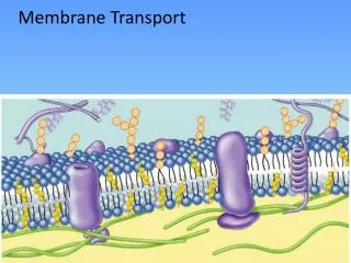
BCOR 011 Lecture 10 Sept 21, 2005 Membrane Transport. Membrane Transport Permeability Diffusion Role of transport proteins - facilitated Channel proteins Carrier proteins 4. Active vs passive transport. 1. Lipid bilayers are selectively permeable.

E N D
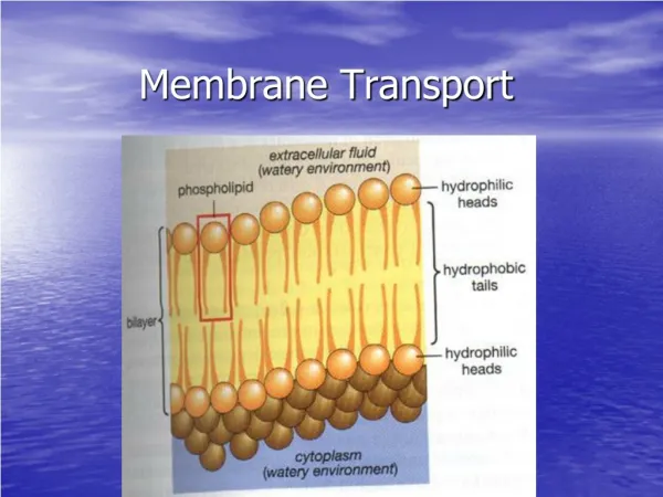
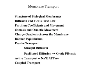
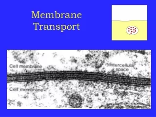
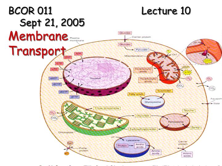
BCOR 011 Lecture 10 Sept 21, 2005 Membrane Transport
Membrane Transport • Permeability • Diffusion • Role of transport proteins - facilitated • Channel proteins • Carrier proteins • 4. Active vs passive transport
1. Lipid bilayers are selectively permeable • small,nonpolar • small • uncharged, polar • larger • uncharged, polar • molecules • ions Decreasing permeability Size – polarity - ions
The Permeability of the Lipid Bilayer • Hydrophobic molecules • Are lipid soluble and can pass through the membrane rapidly • Polar molecules • Do not cross membrane rapidly • Ions • Do not cross the membrane at all
Transport processes Solutes – dissolved ions and small organic molecules i.e., Na+,K+, H+, Ca++, Cl,- sugars, amino acids, nucleotides Three transport processes: a. Simple diffusion – directly thru membrane b. Facilitated diffusion (passive transport) c. Active transport – requires energy Req Carrier prot
Simple Diffusion: • Tendancy of a material to spread out • Always moves toward equilibrium Equilibrium Net diffusion Net diffusion Net diffusion Figure 7.11 B Equilibrium Equilibrium Net diffusion Net diffusion Net diffusion
simple diffusion example: Oxygen crossing red cell membrane HIGH -> low O2 CO2 HCO3- O2 CO2 Lungs O2 CO2 Tissues O2 CO2 HCO3- HCO3- Driving force: concentration gradient Trying to even out concentration
Lower concentration of solute (sugar) Higher concentration of sugar Same concentration of sugar Selectively permeable mem- brane: sugar mole- cules cannot pass through pores, but water molecules can Water molecules cluster around sugar molecules More free water molecules (higher concentration) Fewer free water molecules (lower concentration) Osmosis Water moves from an area of higher free water concentration to an area of lower free water concentration Figure 7.12 H2Otransport: diffusion from area with low [solute] to one with high [solute] Osmosis Diffusion of water Impermeable Solutes
Hypotonic solution Hypertonic solution Isotonic solution (b) Plant cell. Plant cells are turgid (firm) and generally healthiest in a hypotonic environ- ment, where the uptake of water is eventually balanced by the elastic wall pushing back on the cell. (a) Animal cell. An animal cell fares best in an isotonic environ- ment unless it has special adaptations to offset the osmotic uptake or loss of water. H2O H2O H2O H2O H2O H2O H2O H2O Turgid (normal) Flaccid Normal Shriveled Plasmolyzed Lysed Animal cells – pump out ions Plants, bacteria – cell walls Figure 7.13
…but most things are too large or toopolar to cross at reasonable rates using simple diffusion Facilitated diffusion: protein–mediated movement down a gradient Transmembrane transport proteins
Solute Carrier protein (b) A carrier protein alternates between two conformations, moving a solute across the membrane as the shape of the protein changes. The protein can transport the solute in either direction, with the net movement being down the concentration gradient of the solute. Figure 7.15 Transmembrane transport proteins allow selective transport of hydrophilic molecules & ions 1. carrier protein Bind solute, conformational change, release Selective binding “turnstile”
EXTRACELLULAR FLUID Channel protein Solute CYTOPLASM (a) A channel protein (purple) has a channel through which water molecules or a specific solute can pass. Figure 7.15 Transmembrane transport proteins allow selective transport of hydrophilic molecules & ions 2. channel protein aqueous channel hydrophilic pore very rapid selective –size/charge “trap door”
Kinetics of simple vs facilitated Diffusion Gets “saturated” Maximum rate Does Not Get “saturated” v (solute concentration gradient) ->
For CHARGED solutes (ions): net driving force is the electrochemical gradient • has both a concentration + charge component; • Ion gradients can create an electrical voltage gradient across the membrane (membrane potential) + + + + + + + + + + + + + + + + + +++ +++ --- --- --- --- +++ +++ -60 mVolts + + +
Channel Proteins: facilitate passive transport Ion channels: move ionsdown an electrochemical gradient; gated “keys” VoltageLigand Mechanosensitive
Ligand-gated ion channel “Wastebasket model” – step on pedal & lid opens
Ligand-gated example: ligand-gated ion channel “Key” - acetylcholine
Voltage-gated channels + - + + + + + + + - - - - - + - - + - - - Note: channels are passive, facilitated transport systems
Protein ion channels: -are passive, facilitated transport systems -require a membrane protein -typically move ions very rapidly from an area of HIGH concentration to one of lower concentration
Carrier proteins: Transport solute across membrane by binding it on one side, undergoing a conformational change and then releasing it to the other side
Example: Glucose transporter GluT1 : carrier-mediated facilitated diffusion Glucoseout (HIGH)->glucose in (low) outside cell 2. Conformational change 3. Glucose Released- Conformational shift T2 • Glucose binds 2. 1. T1 inside cell T1 3. Glucose + ATP glucose-6-phosphate + ADP hexokinase
Carrier proteins: three types (a) Uniport (b) Co-transport Uniport – onesolute transported [ Symport –twosolutes in thesamedirection Antiport – twosolutes inoppositedirections
Carrier Proteins can mediate either: 1.Passive transport driving force -> concentration/electrochemical gradient OR 2.Active transport against a gradient; unfavorable requires energy input Note: channel proteins mediate only passive transport
Active transport • Carrier protein moves solute AGAINST its concentration gradient • Requires energy, usually in the form of ATP hydorlysis • Or a favorable gradient established by use of ATP
Active transport: Na+K+ Pump (Na+K+ATPase) P P P 3 Na+ out 2 K+ in P ATP!
3 6 2 1 5 4 Na+ binding stimulates phosphorylation by ATP. Cytoplasmic Na+ binds to the sodium-potassium pump. Phosphorylation causes the protein to change its conformation, expelling Na+ to the outside. K+ is released and Na+ sites are receptive again; the cycle repeats. Loss of the phosphate restores the protein’s original conformation. Extracellular K+ binds to the protein, triggering release of the Phosphate group. [Na+] high [K+] low Na+ Na+ Na+ Na+ Na+ ATP [Na+] low [K+] high P Na+ ADP CYTOPLASM The sodium -potassium pump EXTRACELLULAR FLUID Na+ Na+ Na+ K+ P K+ K+ P P i K+ Figure 7.16 K+ K+
The Na+/K+ Pump: Na+ Na+ “bilge pump” Na+ Na+ Na+ Creates an electrochemical gradient (high external [Na+ ]) Na+ potential energy – like “storing water behind a dam” Na+ Na+ Na+ uses ~1/3 of cell’s ATP!!
Example of indirect active transport: Na+ gradient drives other transport Na+ glucosesymport Glucose Gradient Coupled transport
– EXTRACELLULAR FLUID + – ATP + H+ H+ Proton pump H+ + – H+ H+ + – CYTOPLASM + H+ + – • An electrogenic pump • Is a transport protein that generates the voltage across a membrane Figure 7.18
– + H+ ATP H+ + – H+ Proton pump H+ – + H+ – + H+ Diffusion of H+ Sucrose-H+ cotransporter H+ – + – Sucrose + Figure 7.19 • Cotransport: active transport driven by a concentration gradient
Direct active Indirect active transport transport Transport coupled to Exergonic rxn, i.e. ATP hydrolysis *Transport driven by cotransport of ions *note that the favorable ion gradient was established by direct active transport
….Each membrane has its own characteristic set of transporters
Summary: Passive transport Active transport Simple diffusion Facilitated diffusion channel carrier protein protein No protein carrier protein HIGH to low conc low to HIGH conc HIGH to low conc favorable favorable Unfavorable Add energy ATP Figure 7.17