Download
1 / 29
300 likes | 613 Views
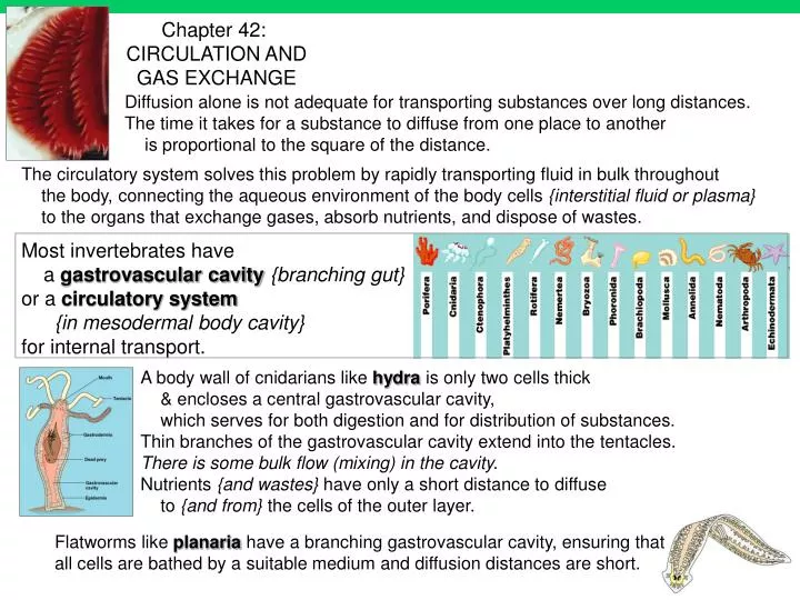
Most invertebrates have a gastrovascular cavity {branching gut} or a circulatory system {in mesodermal body cavity} for internal transport. A body wall of cnidarians like hydra is only two cells thick & encloses a central gastrovascular cavity,

E N D
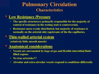
Most invertebrates have a gastrovascular cavity{branching gut} or a circulatory system {in mesodermal body cavity} for internal transport. A body wall of cnidarians like hydra is only two cells thick & encloses a central gastrovascular cavity, which serves for both digestion and for distribution of substances. Thin branches of the gastrovascular cavity extend into the tentacles. There is some bulk flow (mixing) in the cavity. Nutrients {and wastes} have only a short distance to diffuse to {and from} the cells of the outer layer. Flatworms like planaria have a branching gastrovascular cavity, ensuring that all cells are bathed by a suitable medium and diffusion distances are short. Chapter 42: CIRCULATION AND GAS EXCHANGE Diffusion alone is not adequate for transporting substances over long distances. The time it takes for a substance to diffuse from one place to another is proportional to the square of the distance. The circulatory system solves this problem by rapidly transporting fluid in bulk throughout the body, connecting the aqueous environment of the body cells {interstitial fluid or plasma} to the organs that exchange gases, absorb nutrients, and dispose of wastes.
In insects, other arthropods & most mollusks{except cephalopods}, blood bathes the organs directly in an opencirculatory system. There is no distinction between blood and interstitial fluid, and the general body fluid is more correctly termed hemolymph. In insects and other arthropods, the heart is an elongated dorsal tube; it pumps hemolymph through vessels out into sinuses. When the heart relaxes, it draws in hemolymph through pores (ostia). Body movements squeeze the sinuses & help circulate the hemolymph. Instead of lungs, insects have trachea that take air directly to cells. Earthworms, squids, octopuses, and vertebrates have closedcirculatory systems: blood is confined to vessels and is distinct from the interstitial fluid. One or more hearts pump blood into large vessels that branch into smaller ones coursing through the organs, where materials are exchanged by diffusion between the blood and the interstitial fluid bathing the cells. Note: earthworm ‘lungs’ are just skin capillaries. {amphibians & turtles have some gas exchange through skin} For animals with many cell layers, gastrovascular cavities are insufficient. Two types of {mesodermal} circulatory systems have evolved to overcome the limitations of diffusion: open circulatory systems and closed circulatory systems. 3 basic components: a fluid (blood), tubes (blood vessels), a muscular pump (heart).
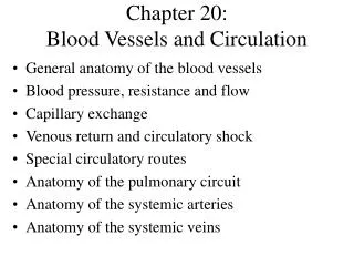
2 3 4 A fish heart has two main chambers, one atrium and one ventricle. Blood pumped from the ventricle travels to the gills, where it picks up oxygen and disposes of carbon dioxide across capillary walls. The gill capillaries converge into a vessel that carries oxygen-rich blood to capillary beds in all other parts of the body (the systemic circulation ). Blood then returns in veins to the atrium of the heart. In fish, blood must pass through two capillary beds during each circuit {like hepatic circulation in mammals} w/ a big drop in pressure across each capillary bed; systemic circulation is quite slow, constraining the delivery of oxygen to body tissues, and hence the maximum aerobic metabolic rate of fishes.
2 3 4 Frogs and other amphibians have a three-chambered heart, with two atria and one ventricle. The ventricle pumps blood into a forked artery that splits the ventricle’s output. The pulmocutaneous circulation leads to capillaries in the gas-exchange organs (the lungs and skin in a frog). Most of the oxygen-rich blood that returns to the ventricle is diverted into the systemic circulation by a ridge in the ventricle. Systemic circulation supplies all body organs and then returns oxygen-poor blood to the right atrium via the veins. This double circulation provides a vigorous flow of blood to the brain, muscles, and other organs because the blood is pumped a second time after it loses pressure in the capillary beds of the lungs or skin. Plethodontid (lungless) salamanders are long & thin; pulmocutaneous circulation is all cutaneous.
2 3 4 Turtles, snakes & lizards {formerly known as Reptiles } also have double circulation with pulmonary (lung) and systemic circuits. Although the ‘reptilian’ heart is three-chambered, the ventricle is partially divided and there is even less mixing of oxygen-rich and oxygen-poor blood than in amphibians. In birds & mammals, the ventricle is completely divided: the left side of the heart receives and pumps only oxygen-rich blood, while the right side handles only oxygen-poor blood. The evolution of double circulation w/ a powerful four-chambered heart was an essential to support the endothermy of birds and mammals. Endotherms use ten times as much energy per gram as ectotherms; their circulatory systems need to deliver about ten times as much fuel and oxygen to their tissues (and remove ten times as much CO2 and other wastes). In the crocodilians (crocodiles and alligators), the ventricle is completely divided into separate right and left chambers. Birds and mammals descended from different reptilian ancestors; their powerful four-chambered hearts evolved independently, an example of convergent evolution.
In the fetus the lungs are nonfunctional and the blood largely bypasses them. As the blood from the inferior vena cava enters the right atrium, a large proportion of it is shunted directly into the left atrium through an opening called the foramen ovale. A small valve, the septum primum, located on the left side of the atrial septum overlies the foramen ovale and helps prevent blood from moving in the reverse direction. The rest of the fetal blood entering the right atrium, passes into the right ventricle and out through the pulmonary trunk. Most of the blood in the pulmonary trunk bypasses the lungs by entering a fetal vessel called the ductus arteriosus which connects the pulmonary trunk to the descending portion of the aortic arch. Fetal heart is functionally 3 chambered, bypassing the lungs {like a salamander!}
Cohort study of multiple brain lesions in sport divers: Role of a patent foramen ovale.Knauth et al. British Medical Journal 314 (7082): 701-705 MAR 8 1997
http://www.americanheart.org/Heart_and_Stroke_A_Z_Guide/athero.htmlhttp://www.americanheart.org/Heart_and_Stroke_A_Z_Guide/athero.html … occlusive coronary atherosclerosis. The coronary at the left is narrowed by 60 to 70%. The coronary at the right is even worse This is a normal coronary artery with no atherosclerosis the lumen can carry as much blood as the myocardium requires. Atherosclerosis: athero (meaning gruel or paste) and sclerosis (hardness). It involves deposits … in the inner lining of an artery. This build-up is called plaque. … atherosclerosis begins because the innermost layer of the artery becomes damaged.
Monocytes {Fig 42.14} initiate plaque formation by adhering to specific binding sites on the endothelium cells. The monocytes then pass through the endothelium … becoming macrophages … scavenger cells that can surround and ingest bacteria, foreign particles, and other cells. macrophages can take up {oxidized}LDL-cholesterol (O-LDL).Once a macrophage has taken up large amounts of cholesterol … it becomes a foam cell … … platelets adhere to the foam cells, and there is … release of growth factors from the platelets … Smooth-muscle cells migrate to the site and secrete collagen, forming a fibrous plaque. http://www.protein.com/healthclaim.nsf/pages/a_process … the prevailing theory is that atherosclerosisbegins with an injury to the inner layer of the artery wall, the endothelium. LDL{‘bad’}-cholesterol molecules accumulate at the injury site, which attracts large white blood cells called monocytes.
Active and passive smoking, risk of carotid chronic infections, and the atherosclerosis - Prospective results from the Bruneck Study.Kiechl et al. 2002. STROKE 33 (9): 2170-2176.Our study provides the first epidemiological evidence that the proatherogenic effects of cigarette smoking are mediated in part by the chronic infections found in smokers. A better understanding of the vascular pathogenetic mechanisms of smoking may offer novel clues for disease prevention … Identification of periodontal pathogens in atheromatous plaques. Preliminary results. Mastragelopulos et al. 2002. CHIRURG 73 (6): 585-591. Recent studies suggest that chronic infections, including those associated with periodontitis, increase the risk for coronary vascular disease. Methods. 34 human specimens … were examined by use of specific oligonucleotide primers … in polymerase chain reaction (PCR) assays. Results. Twenty (59%) of the 34 specimens tested positive for bacterial 16S rDNA. Conclusion. These findings indicate that periodontal pathogens are present in atherosclerotic plaques, where they may play a role in the development and progression of atherosclerosis leading to coronary vascular disease and other clinical sequelae. Is infection a cause or consequence of atherosclerosis?
Antibiotic therapy in coronary heart disease - Where do we currently stand ?Anand V, Gupta S. CARDIOVASCULAR DRUGS AND THERAPY 15 (3): 209-210 MAY 2001Abstract:A casual association between Chlamydia pneumoniae infection and atherosclerosis remains unresolved but plausible. Evidence comes from sero-epidemiological data, pathological specimen examinations, animal models and in vitro experiments. A number of prospective antibiotic intervention trials targeted against C pneumoniae infection in patients with coronary heart disease are now underway. …
http://www.siumed.edu/surgery/cardiothor/clinical/laserrevasc.htmlhttp://www.siumed.edu/surgery/cardiothor/clinical/laserrevasc.html Transmyocardial revascularization (TMR) … employs a laser to place channels through oxygen deprived heart muscle … these channels provide oxygen-rich blood to the deprived areas of the heart. TMR is prescribed for patients for whom bypass surgery and angioplasty are no longer appropriate. … the concept … is modeled after the reptile heart. Unlike the human heart, which is nourished by blood flowing in arteries located on the outside of the heart muscle, the reptile heart is primarily nourished by internal channels that supply blood from the heart chamber into the … heart muscle. In the 1960s, it was conceived that laser could be used to create similar channels in ailing human hearts. It is now general accepted that the laser channels do not stay open, but promote the growth of new blood vessels.
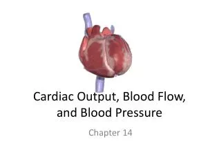
Each day the average heart beats 100,000 times & pumps about 2,000 gallons of blood. In a 70-year lifetime, an average human heart beats more than 2.5 billion times. note: how bundle branches & purkinji fibers reverse direction of contraction: atria top down ventricles bottom up • The normal ECG has the following features: • P wave - due to atrial depolarisation • PR interval (due to delayed conduction through the AV node) • QRS complex - due to ventricular depolarisation • T wave due to ventricular repolarisation
Relate arterial pulses to blood pressure cuff : Contracting skeletal muscles squeeze the veins. Flaps of tissue within the veins act as one-way valves that keep blood moving only toward the heart. If we sit or stand too long, the lack of muscular activity causes our feet to swell with stranded blood. ‘Law of Continuity’ - liquid is incompressible and same volume must pass each cross-section per unit time; ‘shallow rapids & still waters run deep’ Note capillary beds in parallel: if 1 dilates, pressure drops for all & flow redirected to the low-resistance channel Note most pressure drop through Arterioles.
http://www.merck.com/pubs/mmanual/section16/chapter200/200a.htmhttp://www.merck.com/pubs/mmanual/section16/chapter200/200a.htm Orthostatic {Postural} Hypotension An excessive fall in BP (typically > 20/10 mm Hg) on assuming the upright posture. Orthostatic hypotension is not a specific disease but rather a manifestation of abnormal BP regulation due to various causes. Etiology and Pathophysiology The gravitational stress of sudden standing normally causes pooling of blood in the venous capacitance vessels of the legs and trunk. The subsequent transient decrease in venous return and cardiac output results in reduced BP. {and when you stand up, blood pools in your legs, you get dizzy and fall down!} Baroreceptors in the aortic arch and carotid bodies activate autonomic reflexes that rapidly normalize BP by causing a transient tachycardia. {fast heart rate} These changes reflect primarily the sympathetic mediated increase in {neurotransmitter} levels, which augments vasomotor tone of the vessels, increases heart rate and thereby enhances cardiac output {Fig 48.18} ; arterial and venous vasoconstriction are mediated by similar mechanisms. When portions of the autonomic reflex arc are impaired by diseases or drugs, myocardial contractility or vascular responsiveness is depressed, or hypovolemia is present, or hormonal responses are faulty, these homeostatic mechanisms may be inadequate to restore the lowered BP.
Contraction of the smooth muscles in the walls of arterioles and rings of precapillary sphincters controls the flow of blood between arterioles and venules. In anaphylactic shock, a hyper-allergic reaction results in massive release of histamines which constrict bronchiole airways & dilate too many capillaries, dropping blood pressure. Epinephrine should be given to open the airways, and to raise the blood pressure by constricting blood vessels Transfer of substances between the blood and the interstitial fluid occurs only across the thin walls of capillaries. At any given time, only about 5-10% of the body’s capillaries have blood flowing through them. Capillaries in the brain, heart, kidneys, and liver are usually filled to capacity, but in many other sites, like the muscles and gut, the blood supply varies over time as blood is diverted from one destination to another.
Flow follows need. The competition between the parasympathetic and sympathetic nervous systems {Fig 48.18} to direct energy either to storage or to skeletal muscle is also played out in the circulatory system. During times of relaxation, the parasympathetic dominates. blood flow to the visceral organs is relatively abundant. Sympathetic arousal changes the priority of distributing energy in favor of skeletal muscle. Blood vessels to the viscera are constricted and an increased proportion of blood flows to the body wall and limbs. … individual tissues have the ability to signal their own needs for increased blood flow. The messenger is the molecule nitric oxide (NO). NO is synthesized by body tissues in need of oxygen {from arginine} and diffuses across cell membranes. It causes the smooth muscle of the arteriole walls and precapillary sphincters to relax and the vessels to dilate. This interplay of oxygen and NO represents a negative feedback system that can be specific to individual capillary beds and small areas of tissue. http://biology.uindy.edu/Biol504/1review/circulation.htmD
http://www.smartbodyz.com/viagra-yohimbe-arginine-choline-Text.htmhttp://www.smartbodyz.com/viagra-yohimbe-arginine-choline-Text.htm Is That A Bottle In Your Pocket…?Viagra was originally investigated as a potential anti-angina {heart pain} medication, based on its ability to release nitric oxide and increase blood flow to the heart. Although Viagra failed as a heart medication, researchers became excited when many of the men in the clinical trials reported the frequent occurrence of … Vascular smooth muscle (VSM) cells surround arteries and arterioles, contracting and relaxing the arteries to regulate blood pressure. … Normally, in the presence of sexual stimulation, blood flow is directed into … The resulting inflow of blood leads to … This … is triggered by nitric oxide (NO), a short-lived neurotransmitter. Nitric oxide, synthesized from the oxidation of the amino acid arginine, activates an enzyme that manufactures cyclic guanosine monophosphate (cGMP), a biochemical signaling enzyme. cGMP, directs the smooth muscle cells to relax, leading to the dilation of … arteries. However, immediately following release of NO and production of cGMP; another enzyme, cGMP phosphodiesterase type 5 (PDE-5), is activated. PDE-5’s main activity is to destroy cGMP almost as fast as it is formed. The result of this … is a rapid decrease in smooth muscle relaxation and a loss of blood flow … Subsequently, … How Viagra WorksUnfortunately, as we age, cellular concentrations of cGMP decrease. Viagra works … by (1) enhancing the effects of nitric oxide, and (2) maintaining higher levels of cGMP, the two key players in ... The way Viagra does this is to selectively inhibit the cGMP – destroying actions of PDE-5. FDA and Pfizer have warned against taking Viagra with any nitrate-based cardiac medications (i.e., sublingual nitroglycerin tablets, nitroglycerin patches, etc.). There have been cases where patients who received both drugs have died after developing irreversible hypotension
http://www.human-nature.com/darwin/emotion/chap13.htm THE EXPRESSION OF THE EMOTIONS IN MAN AND ANIMALS CHAPTER XIII. Blushing is the most peculiar and the most human of all expressions. Monkeys redden from passion, but it would require an overwhelming amount of evidence to make us believe that any animal could blush. The reddening of the face from a blush is due to the relaxation of the muscular coats of the small arteries, by which the capillaries become filled with blood; and this depends on the proper vaso-motor centre being affected. {then arteriole & sphincter smooth muscle via sympathetic (‘involuntary’) nerves} …as a general rule, with English women, blushing does not extend beneath the neck and upper part of the chest. The small vessels of the face become filled with blood, from the emotion of shame, in almost all the races of man, We have now to consider, why should the thought that others are thinking about us affect our capillary circulation? {consider the idea of an ‘involuntary’ signal from subconscious !}
http://www.merck.com/pubs/mmanual/section22/chapter298/298d.htmhttp://www.merck.com/pubs/mmanual/section22/chapter298/298d.htm Although the brain receives about 1/6 of cardiac output, distribution of drugs to brain tissue is restricted. Lipid soluble substances such as oxygen and carbon dioxide can pass through endothelial cell membranes so can exchange across the entire capillary wall. Some lipid-soluble drugs enter the brain and exert their effects rapidly. Water soluble substancesexchange through many pores {between cells, around tight junctions}. The pores vary in size in different organs. The endothelial cells of the brain capillaries are more tightly joined to one another {more tight junctions} than are those of other capillaries. In the brain not even small ions can leave the capillary by diffusion and there is a blood-brain barrier. Many drugs, particularly the more water-soluble drugs, enter the brain slowly. http://www.sfn.org/briefings/blood-brain.html Researchers are trying to sneak therapies past the barrier to the brain tissue by hitching therapeutic agents onto molecules that already are allowed to slip through or with compounds that pry open the seals.
Blood is a connective tissue with cells suspended in plasma. Plasma is about 90% water w/ many ionic solutes = electrolytes {recall Pedialyte}. Blood normally has a pH of 7.4 in humans. {hyperventillation blows off CO2 & raises pH} The kidney maintains plasma electrolytes at precise concentrations, an example of homeostasis. Various plasma proteins escorts lipids, which are insoluble in water; immunoglobulins or antibodies attack antigens; fibrinogens are clotting factors. Blood plasma that has had these clotting factors removed is called serum.
Two classes of cells are suspended in blood plasma : red blood cells, which transport oxygen, and white blood cells, which function in defense. A third cellular element, platelets, are pieces of cells that are involved in clotting. Mammalian erythrocytes lack mitochondria & nuclei (unusual): more space for hemoglobin. There are five major types of white blood cells; their function is to fight infections {Ch 43}. monocytes & neutrophils are phagocytes; basophils release histamine & eosinophils release peroxidase {chemical weapons}; lymphocytes develop into B cells and T cells, which produce the immune response
Erythrocytes, leukocytes, and platelets all develop from a single population of cells: pluripotent stem cells in the red marrow of bones. "Pluripotent" means these cells have the potential to differentiate into any type of blood cell or into cells that produce platelets {see Ch 21}. Purified pluripotent stem cells may soon provide an effective treatment for a number of human diseases, such as leukemia. A negative-feedback mechanism controls erythrocyte production. If the tissues do not receive enough oxygen, the kidney converts a plasma protein to a hormone called erythropoietin, which stimulates production of erythrocytes. If blood is delivering more oxygen than the tissues can use, the level of erythropoietin is reduced.
Chronic Obstructive Pulmonary Disease Emphysema is characterized by abnormal permanent enlargement of the airspaces distal to the terminal bronchioles with destruction of their walls {greatly reducing S/V & gas exchange} The incidence of emphysema, the fourth leading cause of death in the United States, is up more than 40% since 1982. To obs the recovery of heat & moisture in sinuses: slowly exhale onto wrist via (a) mouth, then (b) nose) The crossing of the airway & ‘foodway’ (trachea & esophagus) is a legacy of the evolution of a bony palate (roof of mouth).
This (& heart valves) is why CPR works
Our breathing control centers in the medulla & pons monitor CO2by slight changes in the pH of the cerebrospinal fluid bathing the brain. (CO2 & H2O form carbonic acid, which lowers the pH.) When the medulla’s control center registers a slight drop in pH (increase in CO2) it increases the depth and rate of breathing, and the excess CO2 is eliminated in exhaled air. O2has little effect on the breathing control centers. However, when the O2 level is severely depressed O2 sensors in the aorta and carotid arteries send alarm signals to the breathing control centers, CO{carbon monoxide}is a lethal poison that is produced when fuels are burned. Because CO is colorless, tasteless, odorless … it can overcome the exposed person without warning. {Why don’t blood gas sensors above give warning?} CO poisons primarily by tightly binding to hemoglobin in the blood {inside the RBCs} replacing oxygen, and reducing the oxygen-carrying capacity of the blood. {but little effect on plasma O2 or CO2 concentrations}