Download
1 / 18
270 likes | 754 Views
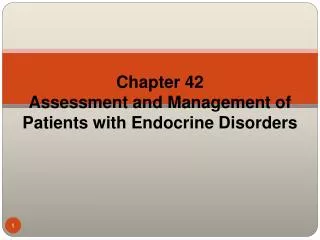
Chapter 16: The Endocrine System. Arnold Adolph Berthold 1803 – 1861 Founder of Endocrinology. Berthold’s Experiment in Roosters…. Castration & Reimplantation of testis. Castration. Castration & Transplantation of testis. Berthold’s Conclusion.

E N D
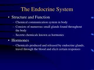
Chapter 16: The Endocrine System
Arnold Adolph Berthold 1803 – 1861 Founder of Endocrinology
Berthold’s Experiment in Roosters…. Castration & Reimplantation of testis Castration Castration & Transplantation of testis
Berthold’s Conclusion... -A secretory, blood-borne product of the transplanted testes is responsible for the normal development of the birds in the second and third group Today, it is called TESTOSTERONE -’problem’: no one knows why Berthold did the experiment in the first place…. No clear rationale for it.
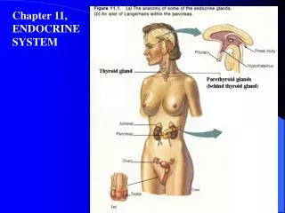
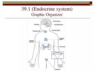
Figure 16.1: Location of the major endocrine organs of the body, p. 605. Pineal gland Hypothalamus Pituitary gland Thyroid gland Parathyroid glands (on dorsal aspect of thyroid gland) Thymus gland Adrenal glands Pancreas Ovary (female) Testis (male)
Figure 16.3: PIP second-messenger mechanism of amino acid-based hormones, p. 608. Extracellular fluid Hormone DAG 1 4 5 Active protein kinase C PIP2 3 2 GTP GTP Receptor Gq Inactive protein kinase C GTP GDP IP3 Phospholipase C Catecholamines TRH ADH GnRH Oxytocin Triggers responses of target cell 5 Endoplasmic reticulum 6 Cytoplasm Ca2+ Ca2+-calmodulin
Figure 16.4: Direct gene activation mechanism of steroid hormones, p. 609. Steroid hormone Cytoplasm Steroid hormone Receptor- chaperonin complex Receptor-hormone complex Molecular chaperones Hormone response elements Binding Transcription Chromatin mRNA mRNA Nucleus New protein Ribosome Translation
Figure 16.5: Three types of endocrine gland stimuli, p. 612. Capillary blood contains low concentration of Ca2+, which stimulates… Preganglionic SNS fiber stimulates adrenal medulla cells… The hypothalamus secretes hormones that… 1 1 1 Hypothalamus Capillary (low Ca2+ in blood) Thyroid gland (posterior view) CNS (spinal cord) …stimulate the anterior pituitary gland to secrete hormones that… 2 Preganglionic SNS fiber Parathyroid glands Pituitary gland Medulla of adrenal gland Thyroid gland Adrenal cortex Gonad (Testis) Parathyroid glands PTH …secretion of parathyroid hormone (PTH) by parathyroid glands 2 Capillary …stimulate other endocrine glands to secrete hormones 3 …to secrete catecholamines 2 (a) Humoral (b) Neural (c) Hormonal
Figure 16.6: Relationships of the pituitary gland and hypothalamus, p. 613. Hypothalamic neurons in the paraventricular nuclei Neurons in the ventral hypothalamus Hypothalamic neurons in the supraoptic nuclei Infundibulum (connecting stalk) Superior hypophyseal artery Hypothalamic- hypophyseal tract Hypophyseal portal system Neurohypophysis (storage area for hypothalamic hormones) • Primary capillary plexus • Hypophyseal portal veins • Secondary capillary plexus Posterior lobe Anterior lobe Secretory cells of adenohypophysis Venule Oxytocin ADH TSH, FSH, LH, ACTH, GH, PRL Inferior hypophyseal artery Venule
Figure 16.7: Metabolic actions of growth hormone (GH), p. 615. Hypothalamus secretes growth hormone – releasing hormone (GHRH), and somatostatin (GHIH) Inhibits GHRH release Stimulates GHIH release Anterior pituitary Feedback mechanism Inhibits GH synthesis and release Key: Growth hormone Increases, stimulates Direct effects Reduces, inhibits Initial stimulus Liver and other tissues Physiological response Result Insulin-like growth factors (IGFs) Anti-insulin actions Indirect growth-promoting actions Extraskeletal effects Carbohydrate metabolism Fat Skeletal effects Increased protein synthesis, and cell growth and proliferation Increased cartilage formation and skeletal growth Increased blood glucose and other anti-insulin effects Increased lipolysis
Figure 16.8: Gross and microscopic anatomy of the thyroid gland, p. 620. Colloid-filled follicles Hyoid bone Follicle cells Epiglottis Thyroid cartilage Internal carotid artery External carotid artery Superior thyroid artery Common carotid artery Isthmus of thyroid gland Inferior thyroid artery Trachea Left subclavian artery Brachiocephalic artery Left lateral lobe of thyroid gland Aorta Parafollicular cell (a) (b)
Figure 16.11: The parathyroid glands, p. 624. Pharynx (posterior aspect) Thyroid gland Capillary Parathyroid glands Chief cells Esophagus Trachea Oxyphil cells (a) (b)
Figure 16.12: Effect of parathyroid hormone on bone, the intestine, and the kidneys, p. 625. Hypocalcemia (low blood calcium) stimulates parathyroid glands Key: = Ca2+ ions = PTH molecules Rising Ca2+ in blood inhibits PTH release PTH release from parathyroid glands PTH: Bone Activates osteoclasts; calcium and phosphate ions released into blood Intestine Increases calcium absorption from food Promotes activation of vitamin D Kidney Increases calcium reabsorption Blood- stream
Figure 16.13: Microscopic structure of the adrenal gland, p. 626. Capsule Zona glomerulosa Adrenal gland Zona fasciculata • Medulla • Cortex Zona reticularis Kidney Adrenal medulla (a) (b)
Figure 16.16: Stress and the adrenal gland, p. 631. Stress Short term More prolonged Hypothalamus CRH (corticotropin- releasing hormone) Nerve impulses Spinal cord Corticotroph cells of anterior pituitary To target in blood Preganglionic sympathetic fibers Adrenal cortex ACTH Adrenal medulla Short-term stress response Mineralocorticoids Glucocorticoids 1. Increased heart rate 2. Increased blood pressure 3. Liver converts glycogen to glucose and releases glucose to blood 4. Dilation of bronchioles 5. Changes in blood flow patterns leading to decreased digestive system activity and reduced urine output 6. Increased metabolic rate Long-term stress response Catecholamines (epinephrine and norepinephrine) 1. Retention of sodium and water by kidneys 2. Increased blood volume and blood pressure 1. Proteins and fats converted to glucose or broken down for energy 2. Increased blood glucose 3. Suppression of immune system
Figure 16.18: Regulation of blood glucose levels by insulin and glucagon, p. 633. Stimulates glucose uptake by cells Insulin Tissue cells Stimulates glycogen formation Pancreas Glycogen Glucose Blood glucose falls to normal range Liver Stimulus: Rising blood glucose level Imbalance Homeostasis: Normal blood glucose level (about 90 mg/100 ml) Stimulus: Declining blood glucose level Imbalance Blood glucose rises to normal range Pancreas Stimulates glycogen breakdown Glucose Glycogen Glucagon Liver
Figure 16.1: Modified to emphasize the relationship between the adrenal glands and the testes and ovaries. Testes & Ovaries These gamete producing glands produce the lion’s share of sex hormone for each sex. Ovaries are in females and Testes are in males. There is, however, an important role for the adrenal glands… Adrenal glands Ovary (female) Testis (male)