Download
1 / 7
70 likes | 185 Views
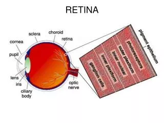
Retina - contains Pigment epithelial cells- absorb light that passes through the retina First neuron Rods- vision in light of low intensity Cones- visual acuity and color vision Second neuron Bipolar- second neurons of visual pathway Ganglion Ganglion- third neurons of visual pathway. Eye

E N D
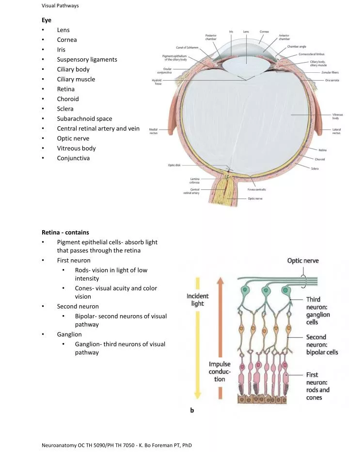
Retina - contains • Pigment epithelial cells- absorb light that passes through the retina • First neuron • Rods- vision in light of low intensity • Cones- visual acuity and color vision • Second neuron • Bipolar- second neurons of visual pathway • Ganglion • Ganglion- third neurons of visual pathway Eye • Lens • Cornea • Iris • Suspensory ligaments • Ciliary body • Ciliary muscle • Retina • Choroid • Sclera • Subarachnoid space • Central retinal artery and vein • Optic nerve • Vitreous body • Conjunctiva Neuroanatomy OC TH 5090/PH TH 7050 - K. Bo Foreman PT, PhD
Visual Pathway- • Light travels through retina from internal to external exciting the rods and cones • Impulse is transferred from rods and cones to bipolar cells • Bipolar cells synapse on the dendrites of the ganglion cells Additional areas: • Optic disc- Area optic nerve exits retina (blind spot- no receptors) • Macula lutea- central region of the retina • Fovea centralis- area of the central part of the retina that is indented and contains mainly cones for visual acuity Neuroanatomy OC TH 5090/PH TH 7050 - K. Bo Foreman PT, PhD
Visual Pathway- forth order neurons • Cell body located in the lateral geniculate body • Axon travels posteriorly (optic radiations) • Terminates in the visual cortex in the occipital lobe Visual Pathway- • Ganglion cells form the optic nerve at the level of the optic disc • Optic nerves combine to form optic chiasm • Enters lateral geniculate body to synapse
1. Right eye blindness - trauma, optic neuritis • 2. Bitemporal hemianopsia - pituitary tumors • 3. Right nasal hemianopsia - pressure by aneurysm of internal carotid artery • 4. Left homonymous hemianopsia - abscess or tumor of temporal lobe • 5. Left homonymous hemianopsia - anterior choroidal artery dysfunction • 6. Left homonymous superior quadratic anopsia - temporal or occipital lobe dysfunction • 7. Left homonymous inferior quadratic anopsia - parietal or occipital lobe tumor • 8. Left homonymous hemianopsia with macular preservation - posterior cerebral artery dysfunctions, tumors, trauma 1. 2. 3. 4. 5. 6. 7. 8. Neuroanatomy OC TH 5090/PH TH 7050 - K. Bo Foreman PT, PhD
Visual Reflexes • Light reflex (constriction- parasympathetic): when light enters the pupil the pupil constricts • Neuronal elements of the retina • Optic nerve • Optic chiasm • Optic tract • Enter the brachium of the superior colliculus (superior brachium) • Pretectal region • Oculomotor nuclear complex (Edinger-Westphal nucleus) • Preganglionic parasympathetic axons to the ciliary ganglion behind eye • Short ciliary nerves • Terminate on the constrictor muscle of the iris Neuroanatomy OC TH 5090/PH TH 7050 - K. Bo Foreman PT, PhD
Visual Reflexes • Light reflex (dilation- sympathetic): when the pupil widens. Occurs passively when parasympathetic tone decreases and actively when sympathetic tone increases • Hypothalamus • Spinal cord (T1) • Ventral root • White communicating rami • Ascend sympathetic trunk • Synapse in superior cervical ganglion with post-synaptic sympathetic ganglion • Travel in the carotid plexus • Terminate on the dilator muscle of the iris Neuroanatomy OC TH 5090/PH TH 7050 - K. Bo Foreman PT, PhD
Visual Reflexes • Accommodation reflex: Process in which a clear visual image is maintained as gaze is shifted from a distant to a near point • 3 components • Convergence of the eyes • Pupillary constriction • Thickening of the lens • Initiated by occipital cortex • Impulses go to: • Edinger-Westphal nucleus for changes in the lens and pupil (parasympathetic) • Somatic nuclei: for convergence of the eyes (medial rectus Neuroanatomy OC TH 5090/PH TH 7050 - K. Bo Foreman PT, PhD