Download
1 / 33
530 likes | 2.13k Views
Equine Hind Limb. Vessels and Nerves Tarsal Radiographs Paragraphs 294-305 March 29, 2000 Dr. Provo. Sacrosciatic Ligament - Nerves (S363). greater sciatic foramen. lesser sciatic foramen. caudal gluteal nerve. caudal rectal nerve. pudendal nerve. caudal cutaneous femoral nerve.

E N D
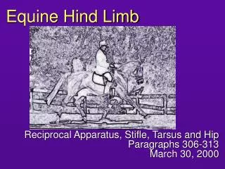
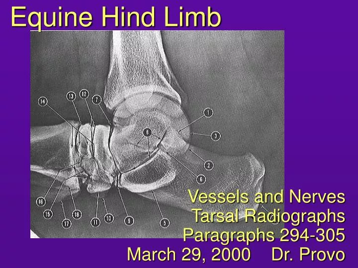
Equine Hind Limb Vessels and Nerves Tarsal Radiographs Paragraphs 294-305 March 29, 2000 Dr. Provo
Sacrosciatic Ligament - Nerves (S363) greater sciatic foramen lesser sciatic foramen caudal gluteal nerve caudal rectal nerve pudendal nerve caudal cutaneous femoral nerve sciatic nerve cranial gluteal nerve ischiatic spine greater trochanter
Vessels Associated with Sacrosciatic Ligament (S683) Lateral view cranial gluteal caudal gluteal internal pudendal anastomotic branch of obturator vein
Motor Nerves of the Lumbosacral Plexus Going to the Hind Limb Nerve Muscles Supplied semimembranosus, semitendinosus, biceps femoris, rotator muscles of hip Sciatic sublumbar muscles, quadriceps femoris, sartorius (saphenous nerve) Femoral Obturator pectineus, gracilis, adductor, external obturator Cr. Gluteal tensor fascia lata, gluteal muscles Cd. Gluteal superficial and middle gluteal, vertebral head of biceps femoris and semitendinosus Common Peroneal biceps femoris, flexors of hock, extensors of digit Tibial extensors of hock, flexors of digit Reminder: REVIEW flexion and extension in the rear limb!
flexion flexion
Thinking ahead to 2002-2003… • A horse has a bump in this location: What joint is in this area? • This is a common injection site: What muscles are involved? • There is a swelling here: What structure could be involved? sacroiliac biceps femoris and semitendinosus popliteal lymph node
Gluteal Muscles: Organization, and Insertion Points Superficial Middle Deep Third trochanter Convexity of greater trochanter Superficial part Deep part (“accessory head”) main part “piriformis” Crest distal to convexity Summit of greater trochanter Intertrochanteric crest and third trochanter Trochanteric bursa
Greater Trochanter Summit (insertion of main part of superficial part of middle gluteal) Area of bursa: Convexity (deep gluteal inserts on medial side) Intertrochanteric crest between greater and third trochanters (insertion of piriformis of superficial part of middle gluteal) Roughened area for insertion of deep part of middle gluteal: third trochanter (insertion of superficial gluteal and of piriformis)
Leg Muscles long digital ext. lat. dig. extensor deep dig. flexor • extensor retinacula: • proximal • middle • distal • lateral S685 S445, lateral view
Extensor Retinacula S447 • Peroneus tertius (yellow) • Cranial tibial (blue) • Long Digital Extensor (green) • Lateral Digital Extensor (red) • Extensor Retinacula (orange) • proximal • lateral (not shown) • middle • distal • Emergence of the cranial tibial tendon from bifurcation of the peroneus tertius M
Cutaneous Innervation: some general notes • Three nerves supply the sensory innervation from mid-crus distally: • Saphenous (a branch of the femoral); tibial; common peroneal (the last two are branches of the sciatic) • There is a lot of overlap between branches of common peroneal and tibial nerves. • General distribution: • Common peroneal to dorsal and dorsolateral surface of hock and metatarsus • Tibial to plantar and plantaromedial surface of hock and metatarsus. • Overlap in the digit and plantarolateral surface of metatarsus.
Cutaneous Innervation 12 = branch of common or superficial peroneal 13 = branch of tibial 12a. lat. cut. sural 11. saphenous 13. caudal cutan. sural 12b. sup. peroneal 12a. med. or lat. dorsal metatarsal n. 13. dorsal br. of digital n.
Example Quiz Questions • What is the action of these muscles on the hock? • What nerve innervates these muscles? flexion common peroneal
tibial common peroneal superficial peroneal lateral plantar deep peroneal medial plantar lat. dors. metatarsal med. dors. metatarsal lat.plant. digital med. plant. digital lat. plant. metatarsal lat. dors. digital med. plant. metatarsal med. dors. digital dorsal branch dorsal branch Sensory Nerve Supply to Distal Limb: Summary sciatic
Schematic of Common Peroneal and Tibial Nerves • Sciatic divides: • Common peroneal • Tibial • Common peroneal divides: • Superficial branch • Deep branch • stays with cranial tibial artery • Tibial divides: • Medial plantar • Lateral plantar Note that the rear digit has both plantar and dorsal nerves. From the Mediclip image bank
Tibial Nerve, Plantar View – branching identical to pattern in forelimb Tib. F M L tibial nerve T C medial plantar nerve lateral plantar nerve c 4 1+2 3 medial plantar metatarsal nerve 2 4 3 lateral plantar metatarsal nerve communicating branch
Deep Branch of Peroneal Nerve (Dorsal View) L M peroneal nerve, deep branch lateral dorsal metatarsal nerve medial dorsal metatarsal nerve (both dorsal metatarsalsexchange fibers with plantar metatarsal nerves)
Digital and Metatarsal Nerves, Medial View medial dorsal metatarsal n. (from deep peroneal) medial plantar (from tibial) medial plantar metatarsal (from tibial) medial dorsal digital nerve (from peroneal and tibial) medial plantar digital nerve dorsal branch of medial plantar digital nerve
femoral popliteal cranial tibial dorsal pedal dorsal metatarsal a. III (great metatarsal a.) (dist. perforating branch) m. & l. digitals Arterial Supply to Distal Limb: Overview external iliac saphenous caudal femoral caudal tibial anastomosis (at hock) perforating tarsal (prox. perf. branch) plantar arteries deep plantar arch plantar metatarsal arteries
Example Quiz Question • What branch of the sciatic nerve supplies the plantar nerves? • The cranial tibial artery continues across the dorsal aspect of the tarsus as the artery. tibial dorsal pedal
From the Mediclip image bank Schematic of Major Arterial Supply ext. iliac femoral popliteal cr. tibial dors. pedal d. metatars. III dist. perf. branch digital aa. palpable medial view (Sack and Habel p. 104)
Origin of (Plantar) Digital Vessels main blood supply from dorsal metatarsal III (great metatarsal a.) crosses between MT III & IV (as distal perforating br.) divides into medial and lateral digital arteries medial and lateral plantar (and plantar metatarsal, not shown) arteries join plantar digitals at fetlock lateral plantar
III II IV IM DDF SDF Cross Section - Metatarsus long digital extensor dorsal metatarsal artery III and lat. dorsal metatarsal nerve lat. plantar metatarsal artery and nerve lat. plantar vein, artery, nerve medial dorsal metatarsal nerve plantar mt. v. med. plantar mt. artery and nerve dorsal common dig v. II med. plantar vein, artery, nerve
General Comments about Digital Vessels and Nerves of the Rear Digit • The arrangement of the digital vessels is essentially identical in the rear digit to that in the thoracic digit. • However, the rear digital vessels have more collateral circulation contributing to their formation than in the forelimb. • The arrangement of the plantar digital nerves is essentially identical to that of the palmar digital nerves. • The rear limb also has dorsal digital nerves. • Remember this when you do a nerve block in the rear digit.
Example Quiz Question 1. Breed? 2. What nerve can be palpated here? 3. What artery can be used to take a pulse here? thoroughbred tibial dorsal MT III
Thinking Ahead to 2002-2003… • Which radiographic projection of the tarsus would be the best to demonstrate each of the following lesions? • Fracture of the lateral trochlear ridge (hint: this is on the dorsolateral aspect of the tarsus…) • Separation of MT II and III • Chip fracture of the lateral aspect of tarsal bone 4 plantarolateral – dorsomedial oblique plantarolateral – dorsomedial oblique dorsoplantar Left tarsus
Dorsoplantar (DPl) Tarsal Radiographs left dorsal right plantar
Lateromedial (LM) Tarsal Radiographs left lateral right medial (slightly oblique)
Oblique Tarsal Radiographs Dorsolateral-Plantaromedial Oblique (DLPM-O) of Left Tarsus Plantarolateral – Dorsomedial Oblique (DMPL-O) of Left Tarsus Usually called a DMPL-O, even though the x-rays went the opposite direction. (They usually don’t put the head of the machine under the horse.) Areas best visualized:
Dorsomedial-Plantarolateral Oblique (DMPL-O) of Left Tarsus Notice the deep notch on the lateral trochlear ridge.
Example Quiz Question 1. Name the radiographic projection shown. 2. Which aspect of the tarsus is this? Dorsolateral-plantaromedial oblique Plantarolateral (hint: you can see the calcaneus on the free edge)