Download
1 / 24

E N D
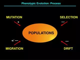
Mechanisms of Spontaneous Mutation • The origin of spontaneous hereditary change has always been a topic of considerable interest. It is known now that spontaneous mutations arise from a variety of sources, including errors in DNA replication, spontaneous lesions, and other more complex mechanisms. • Spontaneous mutations are very rare, making it difficult to determine the underlying mechanisms. How then do we have insight into the processes governing spontaneous mutation? Even though they are rare, some selective systems allow numerous spontaneous mutations to be obtained and then characterized at the molecular level for example, their DNA sequences can be determined. From the nature of the sequence changes, inferences can be made about the processes that have led to the spontaneous mutations.
Errors in DNA replication. • Mispairing in the course of replication is a source of spontaneous base substitution. Tautomers of bases • Each of the bases in DNA can appear in one of several forms, called tautomers, which are isomers that differ in the positions of their atoms and in the bonds between the atoms. The forms are in equilibrium. The keto form of each base is normally present in DNA, whereas the imino and enol forms of the bases are rare. Mispairs resulting from the change of one tautomer into another are termed a tautomeric shift. • Most mispairing mutations are transitions. This is likely to be because an A·C or G·T mispair does not distort the DNA double helix as much as A·G or C·T base pairs do. An error in DNA replication can occur when an illegitimate nucleotide pair (say, AC) forms in DNA synthesis, leading to a base substitution. • Mispairs can also result when one of the bases becomes ionized. This type of mispair may occur more frequently than mispairs due to imino and enol forms of bases.
Base mismatches Mismatched bases. (a) Mispairs resulting from rare tautomeric forms of the pyrimidines; (b) mispairs resulting from rare tautomeric forms of the purines.
From mispairs to mutations (a) A guanine undergoes a tautomeric shift to its rare enol form (G*) at the time of replication. (b) In its enol form, it pairs with thymine. (c and d) In the next replication, the guanine shifts back to its more stable keto form. The thymine incorporated opposite the enol form of guanine, seen in part b, directs the incorporation of adenine in the subsequent replication. The net result is a GCAT mutation. If a guanine undergoes a tautomeric shift from the common keto form to the rare enol form at the time of incorporation (as a nucleoside triphosphate, rather than in the template strand diagrammed here), it will be incorporated opposite thymine in the template strand and cause an AT GC mutation.
Transitions vs transversions Transitions • All the mispairs described so far lead to transitions, in which a purine substitutes for a purine or a pyrimidine for a pyrimidine. The bacterial DNA polymerase III has an editing capacity that recognizes such mismatches and excises them, thus greatly reducing the mutation rate. Other repair systems corrects many of the mismatched bases that escape correction by the polymerase editing function. Transversions • In transversions, a pyrimidine substitutes for a purine or vice versa. Transversions cannot be generated by the mismatches due to tautomeries. With bases in the DNA in the normal orientation, creation of a transversion by a replication error would require, at some point in the course of replication, mispairing of a purine with a purine or a pyrimidine with a pyrimidine. Although the dimensions of the DNA double helix render such mispairs energetically unfavorable, we now know from X-ray diffraction studies that G A pairs, as well as other purine purine pairs, can form.
Frameshift mutations A model for frameshift formation. (a-c) In DNA synthesis, the newly synthesized strand slips, looping out one or several bases. This loop is stabilized by the pairing afforded by the repetitive-sequence unit (the A bases in this case). An addition of one base pair, AT, will result at the next round of replication in this example. (d-f) If, instead of the newly synthesized strand, the template strand slips, then a deletion results. Here the repeating unit is a CT dinucleotide. After slippage, a deletion of two base pairs (CG and TA) would result at the next round of replication.
Spontaneous DNA lesions • In addition to replication errors, spontaneous lesions, naturally occurring damage to the DNA, can generate mutations. Two of the most frequent spontaneous lesions result from depurination and deamination. • Depurination, the more common of the two, consists of the interruption of the glycosidic bond between the base and deoxyribose and the subsequent loss of a guanine or an adenine residue from the DNA. • A mammalian cell spontaneously loses about 10,000 purines from its DNA in a 20-hour cell-generation period at 37°C. If these lesions were to persist, they would result in significant genetic damage because, in replication, the resulting apurinic sites cannot specify a base complementary to the original purine. However, efficient repair systems remove apurinic sites. • The deamination of cytosine yields uracil. Unrepaired uracil residues will pair with adenine in replication, resulting in the conversion of a GC pair into an AT pair (a GCAT transition).
A mutational spectrum The distribution of 140 spontaneous mutations in lacI. Each occurrence of a point mutation is indicated by a box. Red boxes designate fast-reverting mutations. Deletions (gold) are represented below. The I map is given in terms of the amino acid number in the corresponding I-encoded lac repressor. Allele numbers refer to mutations that have been analyzed at the DNA sequence level. The mutations S114 and S58 (circles) result from the insertion of transposable elements. S28 (red circle) is a duplication of 88 base pairs.
Hot spot mutation sites • In the 1970s, Jeffrey Miller and his co-workers examined mutational hot spots in the lacI gene of E. coli.Hot spots are sites in a gene that are much more mutable than other sites. The lacI work showed that certain hot spots result from repeated sequences. In lacI, a four-base-pair sequence repeated three times in tandem in the wild type is the cause of the hot spots (for simplicity, only one strand of the double strand of DNA is indicated): • The major hot spot, represented here by the mutations FS5, FS25, FS45, and FS65, results from the addition of one extra set of the four bases CTGG to one strand of the DNA. This hot spot reverts at a high rate, losing the extra set of four bases. The minor hot spot, represented here by the mutations FS2 and FS84, results from the loss of one set of the four bases CTGG. This mutant does not readily regain the lost set of four base pairs.
Deletions and duplications • Large deletions (more than a few base pairs) constitute a sizable fraction of sponta-neous mutations, as already shown. The majority, although not all, of the deletions occur at repeated sequences. The figure below shows the results for the first 12 deletions analyzed at the DNA sequence level, presented by Miller and his co-workers in 1978. Further studies showed that hot spots for deletions are in the longest repeated sequences. Duplications of segments of DNA have been observed in many organisms. Like deletions, they often occur at sequence repeats. How do deletions and duplications form? Deletions may be generated as replication errors. For example, an extension of the model of slipped mispairing could explain why deletions predominate at short repeated sequences. Alternatively, deletions and duplications could be generated by recombinational mechanisms.
Induced mutations • Mutations are categorized as induced or spontaneous.Induced mutations are defined as those that arise after purposeful treatment with mutagens, environmental agents that are known to increase the rate of mutations. • Spontaneous mutations are those that arise in the absence of known mutagen treatment. They account for the "background rate" of mutation and are the ultimate source of natural genetic variation that is seen in populations.The frequency at which spontaneous mutations occur is low, generally in the range of one cell in 105 to 108. • Therefore, if a large number of mutants is required for genetic analysis, mutations must be induced. The induction of mutations is accomplished by treating cells with mutagens.
Spontaneous vs induced mutations • Recognize that the distinction between induced and spontaneous is purely operational. If we are aware that an organism was mutagenized, then we infer that any mutations that arise after this mutagenesis were induced. However, this is not true in an absolute sense. The mechanisms that give rise to spontaneous mutations also are in action in this mutagenized organism. In reality, there will always be a subset of mutations recovered after mutagenesis that are independent of the action of the mutagen. The proportion of mutations that fall into this subset depends on how potent a mutagen is. The higher the rate of induced mutations, the lower the proportion of recovered mutations that are actually "spontaneous" in origin. • Induced and spontaneous mutations arise by generally different mechanisms. After considering these mechanisms, we shall explore the subject of biological mutation repair. Without these repair mechanisms, the rate of mutation would be so high that cells would accumulate too many mutations to remain viable and capable of reproduction. Thus, the mutational events that do occur are those rare events that have somehow been overlooked or bypassed by the repair processes.
Forward mutation frequencies obtained with various mutagens in Neurospora • The assay measures the frequency of ad-3 mutants. It so happens that such mutants are red, so they can be detected against a background of white ad-3+ colonies.
Specificity of mutagens The distribution of mutations among 36 sites in the lacI gene is shown for three mutagens: EMS, UV light, and aflatoxin B1. The height of each bar represents the number of occurrences of mutations at the respective site. Some hot spots are shown off-scale, with the number of occurrences indicated directly above the respective peak. For instance, in the UV-generated collection, one site resulting from a GCAT transition is represented by 80 occurrences. Each mutational site represented in the figure generates an amber (UAG) codon in the corresponding mRNA. The mutations are arranged according to the type of base substitution. Asterisks mark the positions of 5-methylcytosines.
Still on mutagen specificity • The previous graphs show the distribution of base-substitution mutations that create chain-terminating UAG codons. The specific sequence changes are known for each lacI site, allowing the graphs to be broken down into each category of substitution. • The graphs reveals the two components of mutational specificity. First, each mutagen shown favors a specific category of substitution. • For example, EMS and UV favor GCAT transitions, whereas AFB1 favors GCTA transversions. These preferences are related to the different mechanisms of mutagenesis. • Second, even within the same category, there are large differences in mutation rate. • These differences can be seen best with UV light for the GCAT changes. Some aspect of the surrounding DNA sequence must cause these differences. In some cases, the cause of mutational hot spots can be determined by DNA sequence studies, as previously described for certain frameshift sites. In many examples of mutagen-induced hot spots, the precise reason for the high mutability of specific sites is still unknown.
Mechanisms of mutagenesis • Mutagens induce mutations by at least three different mechanisms. • They can replace a base in the DNA • They can alter a base so that it specifically mispairs with another base, • They can damage a base so that it can no longer pair with any base under normal conditions.
Incorporation of base analogs • Some chemical compounds are sufficiently similar to the normal nitrogen bases of DNA that they occasionally are incorporated into DNA in place of normal bases; such compounds are called base analogs. Once in place, these analogs have pairing properties unlike those of the normal bases; thus, they can produce mutations by causing incorrect nucleotides to be inserted opposite them in replication. The original base analog exists in only a single strand, but it can cause a nucleotide-pair substitution that is replicated in all DNA copies descended from the original strand. • For example, 5-bromouracil (5-BU) is an analog of thymine that has bromine at the C-5 position in place of the CH3 group found in thymine. This change does not affect the atoms that take part in hydrogen bonding in base pairing, but the presence of the bromine significantly alters the distribution of electrons in the base. The normal structure (the keto form) of 5-BU pairs with adenine, as shown here. 5-BU can frequently change to either the enol form or an ionized form; the latter pairs in vivo with guanine.
Specific mispairing • Some mutagens are not incorporated into the DNA but instead alter a base, causing specific mispairing. Certain alkylating agents, such as ethylmethanesulfonate (EMS) and the widely used nitrosoguanidine (NG), operate by this pathway: Although such agents add alkyl groups (an ethyl group in EMS and a methyl group in NG) to many positions on all four bases, mutagenicity is best correlated with an addition to the oxygen at the 6 position of guanine to create an O-6-alkylguanine. This addition leads to direct mispairing with thymine, and would result in GCAT transitions at the next round of replication. As expected, determinations of mutagenic specificity for EMS and NG show a strong preference for GCAT transitions.
Intercalating agents • The intercalating agents form another important class of DNA modifiers. These agents are planar molecules, which mimic base pairs and are able to slip themselves in (intercalate) between the stacked nitrogen bases at the core of the DNA double helix (see figure). In this intercalated position, the agent can cause single-nucleotide-pair insertions or deletions. Intercalating agents may also stack between bases in single-stranded DNA.
Base damage and SOS repair • A large number of mutagens damage one or more bases, so no specific base pairing is possible. The result is a replication block, because DNA synthesis will not proceed past a base that cannot specify its complementary partner by hydrogen bonding. In bacterial cells, such replication blocks can be bypassed by inserting nonspecific bases. The process requires the activation of a special system, the SOS system. The name SOS comes from the idea that this system is induced as an emergency response to prevent cell death in the presence of significant DNA damage. SOS induction is a last resort, allowing the cell to trade death for a certain level of mutagenesis. The SOS system. DNA polymerase III, shown in blue, stops at a noncoding lesion, such as the T C photodimer shown here, generating single-stranded regions that attract the Ssb protein (dark purple) and RecA (light purple), which forms filaments. The presence of RecA filaments helps to signal the cell to synthesize UmuD (red circles), which is cleaved by RecA to yield UmuD (pink circles) and UmuC (yellow ovals). The UmuC is recruited to form a complex with UmuD that permits DNA polymerization to proceed past the blocking lesion.
SOS repair and cancerogenicity • Exactly how the SOS bypass system functions is not clear, although in E. coli it is known to be dependent on at least three genes, recA (which also has a role in general recombination), umuC, and umuD. Current models for SOS bypass suggest that the UmuC and UmuD proteins combine with the polymerase III DNA replication complex to loosen its otherwise strict specificity and permit replication past noncoding lesions. • The previous figure shows a model for the bypass system operating after DNA polymerase III stalls at a type of damage called a TC photodimer. Because replication can restart downstream from the dimer, a single-stranded region of DNA is generated. This region attracts the stabilizing proteinSsb, as well as the RecA protein, which forms filaments and signals the cell to synthesize the UmuC and UmuD proteins. Thiscomplex allows DNA polymerization to continue past the dimer, adding bases across from the dimer with a high error frequency. • Therefore mutagens that damage specific base-pairing sites are dependent on the SOS system for their action. The category of SOS-dependent mutagens is important, because it includes most cancer-causing agents (carcinogens), such as ultraviolet light, aflatoxin B1, and benzo(a)pyrene (discussed later).
Ultraviolet light • Ultraviolet light generates two different lesions that occur at adjacent pyrimidine residues: the cyclobutane pyrimidine photodimer and the 6-4 photoproduct. These lesions interfere with normal base pairing; hence, induction of the SOS system is required for mutagenesis. The insertion of incorrect bases across from UV photoproducts is at the 3’ position of the dimer, and more frequently for 5’-CC-3’ and 5’-TC-3’ dimers. The C T transition is the most frequent mutation, but other base substitutions (transversions) and frameshifts also are stimulated by UV light, as are duplications and deletions. (a) Structure of a cyclobutane pyrimidine dimer. Ultraviolet light stimulates the formation of a four-membered cyclobutane ring (green) between two adjacent pyrimidines on the same DNA strand by acting on the 5,6 double bonds. (b) Structure of the 6-4 photoproduct. The structure forms most prevalently with 5’-CC-3’ and 5’-TC-3’, between the C-6 and the C-4 positions of two adjacent pyrimidines, causing a significant perturbation in local structure of the double helix.
Ionizing radiation • Ionizing radiation results in the formation of ionized and excited molecules that can cause damage to cellular components and to DNA. Because of the aqueous nature of biological systems, the molecules generated by the effects of ionizing radiation on water produce the most damage. Many different types of reactive oxygen specials are produced, including superoxide radicals, such as ·OH. The most biologically relevant reaction products are ·OH, O2, and H2O2. These species can damage bases and cause different adducts and degradation products. Among the most prevalent, which result in mutations, are thymine glycol and 8-oxodG. Ionizing radiation can cause breakage of the N-glycosydic bond, leading to the formation of AP sites, and can cause strand breaks that are responsible for most of the lethal effects of such radiation. DNA damage products formed after attack by oxygen radicals. dR = deoxyribose
Bulky addition products • Aflatoxin B1(AFB1)is a powerful carcinogen, that may contaminate human food. It generates apurinic sites following the formation of an addition product at the N-7 position of guanine. Studies with apurinic sites generated in vitro demonstrated a requirement for the SOS system and showed that the SOS bypass of these sites leads to the preferential insertion of an adenine across from an apurinic site. Thus agents that cause depurination at guanine residues should preferentially induce GC TA transversions. • AFB1 is a member of a class of chemical carcinogens known as bulky addition products when they bind covalently to DNA. Other examples include the diol epoxides of benzo(a)pyrene, a compound produced by internal combustion engines. For many different compounds, it is not yet clear which DNA addition products play the principal role in mutagenesis. In some cases, the mutagenic specificity suggests that depurination may be an intermediate step in mutagenesis; in others, the question of which mechanism is operating is completely open.