Download
1 / 94
950 likes | 1.32k Views
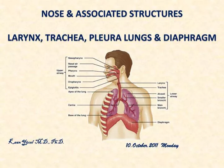
nose & assocIated structures Larynx , trachea , pleura lungs & dIaphragm. Kaan Yücel M.D., Ph.D . 10.October.20 1 1 Monday. nose & assocIated structures. Nose is divisible into two parts as external nose and nasal cavity . Functions of the nose and the nasal cavities are :

E N D
nose& assocIatedstructures Larynx, trachea, pleuralungs & dIaphragm Kaan Yücel M.D., Ph.D. 10.October.2011 Monday
nose & assocIatedstructures • Nose is divisibleintotwoparts as externalnoseandnasalcavity. • Functionsof thenoseandthenasalcavitiesare: • Olfaction (sense of smell) • Respiration • Filtration of thedust in theinspiredair • Humidificationandwarming of theinspiredair • Reception of thesecretionsfromtheparanasalsinusesandnasolacrimalducts
ExternalNosehas fiveparts: • Dorsum • Root • Apex • Nares (nostrils, anteriornasalapertures) • Alae of thenose • External nose has bony and cartilaginous parts.
Bones contributing to the structure of the external nose: • Nasal bones • Frontal process of maxilla • Nasal part of frontal bone
Cartilages contributing to the structure of the external nose: • Lateral cartilages (paired) • Alar cartilages (paired) • Septal cartilage (single)
NasalCavities: • Thetwonasalcavitiesaretheuppermostparts of therespiratorytract. • Theycontaintheolfactoryreceptors. • Thenasalcavitiesareseparated: • fromeachotherby a midlinenasalseptum • fromthe oral cavitybelowbythehard palate • fromthecranialcavityabovebyparts of thefrontal, ethmoid, andsphenoidbones.
Nasal septum is composed of three structures: • Perpendicular plate of the ethmoid bone • Vomer • Septal cartilage
Eachnasalcavity is dividedintoolfactoryarea (upper 1/3) andrespiratoryarea (lower2/3). • Posteriorly, each nasal cavity communicates with the nasopharynx through two openings calledchoana.
Walls of the nasal cavity • Roof of the nasal cavity (anterior to posterior) • nasal bone • frontal bone • cribriform plate of the ethmoid bone • body of the sphenoid bone
Floor of the nasal cavityis formedbythehard palate (palatine process of maxilla and horizontal plate of the palatine bone).
Lateral wall of the nasal cavity (anterior to posterior) • frontal process of maxilla • lacrimal bone • superior nasal concha (of the ethmoid bone), middle nasal concha (of the ethmoid bone), inferior nasal concha • perpendicular plate of the palatine bone • medial lamina of the pterygoidprocess
Medial wall of the nasal cavity is formed by the nasal septum. Themedialwall has a smoothsurface, whereasthelateralwall is unevenduetotheexistance of thenasalconchae.
The spaces between the nasal conchae and the lateral wall of the nasal cavity are called the meatus. • Superior nasal meatus • Middle nasal meatus • Inferior nasal meatus
The following sinuses open into the middle nasal meatus • Frontalsinus • Maxillarysinus • Ethmoid air cells • Sphenoidsinusopensintothesphenoethmoidrecess • Nasolacrimal ductopensintotheinferior nasal meatus
Arterialsupply of thenose The nose has an extensive arterial supply. The branches of the maxillary, ophthalmic and facial arteries supply the nose. Veins of thenose There is a rich network of veinsdeeptothemucosa of thenose. Thisvenous network is important in warmingtheairbefore it entersthetracheaandthelungs. Theveins of thenosedrainintofacialandopthalimicveins.
Nerves of thenose Sensory innervation of the nose is mainly from themaxillarynerve and the ophthalmic nerve. Withintheepithelium of theolfactoryregionliestheolfactorycells (neurons). Theperipheralprocesses of thesecellsterminateunderthemucosaandaresensitivetoodourmolecules in theair. Thecentralprocessesformstheolfactorynerves (CN I).Olfactorynervespassthroughthecribriformplate of theethmoid bone toreachtotheolfactorybulb.
Paranasalsinuses • Paranasal sinuses are air filled spaces lying within the bones around the nasal cavity. • The paranasal sinuses develop as outgrowths from the nasal cavities and erode into the surrounding bones. • All are: • lined by respiratory mucosa, which is ciliated and mucus secreting; • open into the nasal cavities; and • innervated by branches of the trigeminal nerve [V].
Sinuses are named according to the bones they are located in: • Frontal sinuses • Ethmoid sinuses • Sphenoid sinuses • Maxillary sinuses
Frontal sinus • Thefrontalsinuses, one on eachside, arevariable in size andarethemostsuperior of thesinuses. • Thefrontalsinuslies within the inner and outer plates of the frontal bone, posterior to the supercilliary arches and the root of the nose. • The opening of the sinus is called the frontonasal duct. • It drains into the middle nasal meatus.
Ethmoidsinuses Several ethmoid air cells (3-15) collectively are called the ethmoid sinuses. Ethmoid air cells form three groups: Anterior group ------opensintothemiddle meatus Middle group --------opensintothemiddle meatus Posterior group -----opensintothesuperior meatus
Sphenoid sinus • The sphenoid sinus is situated within the body of the sphenoid bone. • Sinusesof eachside is seperatedby a bonyseptum. • Itdrains into the sphenoethmoidal recess.
Maxillary sinus • Themaxillarysinuses, one on eachside, arethelargest of theparanasalsinuses. • Theycompletelyfillthebodies of themaxillae. • The maxillary opening drains into the middle nasal meatus through the semilunar hiatus.
Larynx • Larnynxis the organ of phonation (vocalization). • It is formed of cartilage, muscles and connective tissue. • Larynx’sinnersurface is coveredbytherespiratorymucosa.
Thecavity of thelarynx iscontinuousbelowwiththetrachea, andaboveopensintothepharynximmediatelyposteriorandslightlyinferiortothetongueandtheposterioropening (oropharyngealisthmus) of the oral cavity.
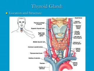
Skeleton of larynxis formed of 3 unpaired and 3 paired cartilages • Unpaired cartilages • Thyroid cartilage (biggest) • Cricoid cartilage • Epiglottic cartilage • Paired cartilages • Arytenoid • Corniculate • Cuneiform
Thyroid cartilage • The thyroid cartilage is the largest cartilage of the larynx. • It is formed of two laminae which fuse anteriorly at the thyroid angleto form laryngeal prominence(Adam’s apple).
Cricoidcartilage • Thecricoidcartilage is a ring shaped cartilage. • Inferiorly it attaches to the first tracheal ring by the cricotracheal ligament.
Arytenoidcartilages • The arytenoid cartilages are pyramidal in shape. • An arytenoid cartilage has three processes: • Apex (superior) • Vocal process (anterior) vocal ligament attaches here • Muscular process (lateral)
Epiglotticcartilage (Epiglottis) • Theepiglottis is a heart shaped cartilage. • Its inferior end is attached to the thyroid cartilage by the thyroepiglottic ligament. • Mostsuperiorend is free.
Corniculateandcuneiform cartilages Theseare small cartilages lying in the posterior part of the aryepiglottic fold.
Fibroelasticmembrane of thelarynx • It lies under the mucosa of the larynx. • The fibroelastic membrane of the larynx has thickenings at certain regions and forms some of the ligaments between the cartilages. • It is formed of two parts: • Quadrangular membrane • Conus elasticus
Conuselasticus (cricovocal membrane):Itsfree upper margin thickens to form the vocal ligament, which is covered by mucosa to form the vocal fold. The opening between the two vocal folds is called rimaglottis. Vocalcord= Vocalfold
The vocal folds are sometimes called 'true vocal folds' to distinguish them from the false vocal folds. These are a pair of thick folds of mucous membrane that protect and sit slightly superior to the more delicate true folds. They have a minimal role in normal phonation, but are often used to produce deep sonorous tones,as well as in musical screaming and the death growl vocal style. The false folds are also called vestibular folds and ventricular folds.
Eachvocal ligament, convergesanteriorly andattaches to the anterior part of the inner surface of the thyroid cartilage (thyroid angle). • Posteriorly, theyindividuallyattach to the vocal processes of the arytenoid cartilages.
Rimaglottis widens during inspiration and two vocal folds are approximated during phonation. • Various changes of the vocal folds determine the color, pitch and the tones of sound. • Pitch increases with tensing, decreasesbyrelaxation. • Intensity of expiration determines the loudness of sound.
Laryngeal Muscles • Extrisic muscles • Thesearethe suprahyoid and infrahyoid muscles. • Theyeither depress or elavate the larynx and hyoid bone. • Intrinsic muscles: There aresixintrinsicmuscles in thelarnyx. Theymovethe laryngeal parts.
INTRINSIC MUSCLES Cricothyroidmuscle; tenses the vocal folds by pulling the thyroid cartilage anteroinferiorly (toproducehigh pitch sound).
Posterior cricoarytenoid muscle; is the only muscle widening the rima glottis.
Lateral cricoarytenoidmuscle;approximates the vocal processes thus, narrowing the anterior part of rima glottis (contracts alone during whispering). Thismuscleadductsthe arytenoid cartilages to close the rimaglottis.
Thyroarytenoid muscle; relaxes the vocal folds by pulling the arytenoid cartilages anteriorly (toproducelowpitchsounds).
Nerves of the larynx Larynx is innervated by the inferiorandsuperiorlaryngealnerves. Boththeinferiorandsuperiorlaryngealnervesarebranches of the vagusnerve (CN X). All of the intrinsic muscles are innervated by the inferior (recurrent) laryngeal nerveexcept the cricothyroid muscle, which is innervated by the external laryngeal nerve(branch of the superior laryngeal nerve).
Trachea • The trachea extends from the inferior end of larynx to the level of T5-T6vertebra. • Itterminatesby dividing into right and left main bronchi.