Download
1 / 46
470 likes | 819 Views
Chronic Rhinosinusitis in Children. Clinical Presentation Hector Stone-Aguilar, M.D. Pediatric Allergy & Immunology Hospital San Jose de Hermosillo Universidad del Valle de Mexico. Clinical presentation of CRS in Children. The problem :.

E N D
Chronic Rhinosinusitis in Children ClinicalPresentation Hector Stone-Aguilar, M.D. PediatricAllergy & Immunology Hospital San Jose de Hermosillo Universidad del Valle de Mexico
Clinical presentation of CRS in Children The problem: • To fully define chronic sinusitis has been difficult • There is a wide variation in clinical expression of the disease • Discordance between patient symptoms and objective findings • No one set of diagnostic criteria has been agreed on by all specialty groups
Clinical presentation of CRS in Children The problem: • Clinical criteria to diagnose CRS, as well as the predictive value of these criteria, are not well defined, specially in children • Historically, the diagnosis of CRS was based on several clinical symptoms, similar to acute RS, but usually less severe • However, none of these symptoms are specific to sinusitis
Definition of Sinusitis • Inflammation of 1 or more of the paranasal sinuses • Acute Sinusitis: less than 4 weeks/duration • Subacute Sinusitis: 4 to 12 weeks/duration • Chronic Sinusitis: longer than 12 weeks Some guidelines also requiring : • Failure to respond to treatment • One positive imaging study Dykewicz M, JACI Feb 03
Definition of Rhinosinusitis • Inflammation of the nose and paranasal sinuses characterized by two or more symptoms, one of which should be either nasal blockage/obstruction/congestion or nasal discharge (anterior/posterior nasal drip) ± facial pain/pressure ± reduction or lost of smell EPOS Guidelines, Rhinology 2007
Rhinosinusitis OHNS , 1997
Definition of Chronic Rhinosinusitis • More than 12 weeks of symptomswithout complete resolution • Can be subdivided in: • ChronicRhinosinusitiswith Nasal Polyps • ChronicRhinosinusitiswithout Nasal Polyps • CRS alsomay be susceptible toexacerbations EPOS Guidelines, Rhinology 2007
CRS: Symptom-based Diagnosis • 73.15% of the nonallergic patients with symptom based diagnosed CRS • 65.34% of the allergic patients with symptom-based diagnosed CRS Had No CT and endoscopic pathology (Endoscopic score 0 + CT score 0) Tahamiler R, Allergy 2007
Chronic Rhinosinusitis in Children In general : The main symptoms associated with rhinosinusitis in children are rhinorrhea, nasal obstruction, mouth breathing, hyponasal speech, and snoring but…
Diagnosing CRS in Children: Special issues Infants and Pre-schoolchildren • Signs/symptomsdifficulttoevaluate: • Congestion (verysubjective/indirect/parent’sbiass) • Only anterior rhinorrheaisreported • Symptomsimpossibletoevaluate: • Posterior discharge • Sense of smell • Headache / toothache / facial pain • Symptomsveryunspecific : • Cough, irritability, fever, fatigue/decreasedactivity, etc.
Diagnosing CRS in Children: Special issues Infants and Pre-schoolchildren • Anterior Rhinoscopy : Limited data • Anterior third of nasal cavity • Osteomeatalzonedifficulttoreach, even w/use of topicaldecongestant • Nasal Endoscopy: Ideal butimpossibletoperformwithoutsedationoranesthesia • CT scan: Also requieres sedationoranesthesia • Sedation/anesthesia: increasescosts and risks • Increasedvalue of plain X-Rays at thisage ??
Severity of Sinusitis • Diseaseseverity can be dividedinto: • Mild (0-3 points) • Moderate (4-7 points) • Severe (8-10 points) • Using a 10-point scoringsystemor Visual AnalogueScale (VAS) EPOS Guidelines, Rhinology 2007
Clinical presentation of CRS in Children Diagnosis must be based in a combination of: • Clinical symptoms and evolution • Age-group related • Previous treatments (type and duration) • Likelihood of allergy involvement: Family history, allergy stigmata, personal history of other allergic diseases (AD or Asthma) • Clinical Signs • Anterior rhinoscopy and/or Nasal endoscopy • Imaging support • Plain X-Rays • CT scans • MRI
Chronic Rhinosinusitis in Children • By definition, needs to be at least 12 weeks old (3 m.o.) • Ethmoid and maxillary sinuses present at birth • Clinical presentation strongly related to the specific pediatric age group: • Infants: Persistent or recurrent rhinorrhea after an acute febrile URIs ( ± AOM, Rhinopharyngitis, Bronchitis) • Pre-schoolars: Persistent rhinorrhea and nasal congestion w/adenoid and tonsil hypertrophy, serous OM, allergies and asthma. • Scholars and adolescents : Nasal obstruction, headaches, sore throath, hyposmia, irritability, sleep disturbances, etc. (PAR or PNAR)
Clinical presentation of CRS in Children • In infants and preschool childrens, most cases of CRS are a chronologic extension of acute infectious sinusitis (viral bacterial) • In contrast, in older children or adolescents most CRS cases are not an infectious disease but an inflammatory disease, much akin to asthma. Jones NS, Curr Opinion Pulm Med, 2000
When to suspect CRS in INFANTS • Continuous or intermittent RHINORRHEA • Anterior, posterior or both • Usually clear initially (days or weeks) • Colored (greenish or yellowish) more dense secretions • It can alternate clear and colored secretions • Nasal CONGESTION • Mild at the beginning • Worsening in an intermittent pattern in absence of appropriate treatment • Not as bad as other age groups • Objective findings: mouth breathing, snoring
When to suspect CRS in INFANTS • COUGH : • A prominent feature of sinusitis at this age • Starts as “Dry” cough usually for several days • Can continue with “wet” cough all the way • Intermittent along the day, not very intense • Can start or worse at night or bedtime • Usually associated with posterior rhinorrhea • Also associated with coarse and audible ronchi • Maybe a better predictor than rhinorrhea about the outcome
When to suspect CRS in INFANTS • FEVER: • Usually present at the beginning of the clinical picture • Low or mid grade • Fades away after few days (with or without treatment) • Can not be present at all • Can relapse in the course of the disease (worsening) • Its absence doesn’t rule out the possibility of chronic infection
When to suspect CRS in INFANTS • Other possible symptoms: • Irritability • Bad appetite • Sleep disturbances: • Trouble to got sleep • Restless sleeping • Nocturnal awakenings • Halitosis • Reduced general activity
When to suspect CRS in INFANTS • Physical signs, NASAL : • Rhinorrhea (anterior) • Pale and enlarged turbinates • Mucosal edema • Hyperemic mucosa • Middle meatus colored discharge
Muco-purulent discharge in the Sinus Ostium zone Middle turbinate Lateral nasal wall Purulent mucus Septum
When to suspect CRS in INFANTS • Physical signs, GENERAL : • Posterior rhinorrhea • Mouth breathing • Pallor • Dark infra-orbital shiners • Halitosis • Tympanic opacity, retraction or hyperemia • Enlarged tonsils • Granular (cobblestone) adenoid tissue in the pharynx • “rude” breathing • Coarse rhonchi on chest examination
Chronic Rhinosinusitis in PRE-SCHOLARS • Not necessarily associated to respiratory infection • Mostly related to allergies and asthma • Difficult to distinguish from PAR. Same sort of signs and symptoms • Usually considered a “complication” of allergic rhinitis • Nasal or sinusal polyps not frequent at this age
Chronic Rhinosinusitis in PRE-SCHOLARS Differences with CRS in Infants • Congestion more prominent than rhinorrhea • Cough frequently related to asthma or BHR • Headaches, frequently mild or intermittent • Hyposmia rarely reported • Halitosis • Clear or thick mucoid rhinorrhea • Paler and more enlarged turbinates • Intense edema of nasal mucosa
Chronic Rhinosinusitis in School children and adolescents • Moderate to severe nasal congestion/obstruction: • Snoring • Sleeping problems • Dry mouth and sore throat at mornings • Headaches: • Mild to severe • Frequent or intermittent • Frontal, maxillary or occipital • Rhinorrhea: • Posterior > anterior • Halitosis
Chronic Rhinosinusitis in School children and adolescents • Daytime somnolence • Tiredness • Poor concentration: altered school performance • Hyposmia • Dysgeusia • Middle ear: • Hypoacusia, Popping, Buzzing • Polyps: More frequent than the other pediatric groups
Consequences of chronic nasal congestion • Snoring • Oral breathing • Hyponasal speech • Sleep disturbances • Obstructive Sleep Apneas (OSA) • Dry mouth • Sore throath • Headaches • Daytime somnolence • Poor concentration • Tiredness • Facial and dental changes
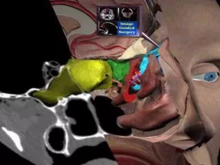
Plain X-rays vs. CT scan in Sinusitis • The sensitivity of Plain X-Ray compared to CT was: • 77% (30/39) • The specificity of the radiograph to CT was 81% (25/31). • The positive likelihood ratio is 4.05 and • The negative likelihood ratio is 0.28. • Conclusions - The difference between radiographs and CT for diagnosing sinus disease in this population is relatively small but favors CT exam. Garcia, DP Radiographic imaging studies in pediatric chronic sinusitis J Allergy Clin Immunol, 94:523-530, 1994.
‘Limited’ CT Scan Garcia D, JACI sept 1994
Sinusitis severity Index (grading):(Glicklich) • Grade 0: mucosal thickening of ≤ 2 mm in any sinusal wall • Grade 1: Any unilateral disease or abnormality • Grade 2: Bilateral disease limited to ethmoid or maxillary sinuses • Grade 3: Bilateral disease with frontal or sphenoidal involvement (any) • Grade 4:Pansinusitis. Emmanuel IA, Otolaryngology Head Neck Surg 2000
CRS Diagnosis:CT scan: Gold standard ? HWANG et al, OHNS april, 2003
CRS Diagnosis:CT scan: Gold standard ? Unilateral involvement of the right maxillary sinus and structural abnormalities: MT concha bullosa and paradoxical curvature of middle turbinate, stretching the OMC
Clasification of the severity of polyposis by endoscopy • 0 - No visible polyps • 1 - Polypsconfinedtothemiddlemeatus • 2 - Polypsbeyondmiddlemeatusbutdid notoccludethe nasal cavity • 3 - Polypsobstructingcompletelythe nasal cavity Mackay IS y Lund VJ, 1997
Nasal / Sinusal Polyposis in Children • If nasal polyps are present in youngchildren, MUST rule out: • AspirinExacerbatedRespiratoryDisease (AERD) • Cystic Fibrosis (CF) • Geneticinvolvement • ButstillmostprobablyrelatedtoPerennialorPersistentAllergicRhinitis • PolypsrelatedtoPerennial Non-AllergicRhinitis are rare at thisage
Etiology of CRS in Children • Infection: • Viral/Bacterial • Biofilms • Fungal? • Allergy • AllergicRhinitis: Persistent > Intermittent • GastroesophagealReflux • Obstruction /Structural • Adenoid > TonsilsHypertrophy • Septaldeviation • Other: concha bullosa, Hallercells, aggernasicells
Etiology of CRS in Children • Immunodeficiency • IgAdeficiency • TransientHipogammaglobulinemia • IgG sub-classdeficiency ( IgG2 + IgG4) • Selective(polysaccaride) IgGdeficiencies • CVI • Cystic Fibrosis • CiliaryDyskinesia • AspirinExacerbatedRespiratoryDisease • Other: veryuncommon
Conclusions: • CRS isfrequent in children • No one set of diagnostic criteria has been agreed on by all specialty groups • CRS in childrenhavespecialfeaturesthat are different of CRS in adultpopulation • There are differencesalso in theclinicalpresentation of thedifferentpediatricagegroups • The diagnosis of CRS in childrenisbasedalmostexclusively in clinical data. Use CT orendoscopy in selected cases. • There are veryfewcontrolledclinicalstudies of CRS in children. AllGuidelinesbased in adultstudies and transpolatedtochildren. • Themostcommon causes are bacterialinfections and/orallergies. Other causes are reallynotfrecuentorrare, butstillhaveto rule outthemifnotresponsive