Download
1 / 5
50 likes | 153 Views
Supplementary Fig. S1. Ct. 0. 5. 10. 15. 20. 0. -0.5. -1. ○. -1.5. -2. ○. log DNA ( pmol). -2.5. -3. ○. -3.5. -4. -4.5. P1. P2. ○. Standard curve of the PCR amplification efficiency of transcripts from the P1 and P2 promoters

E N D
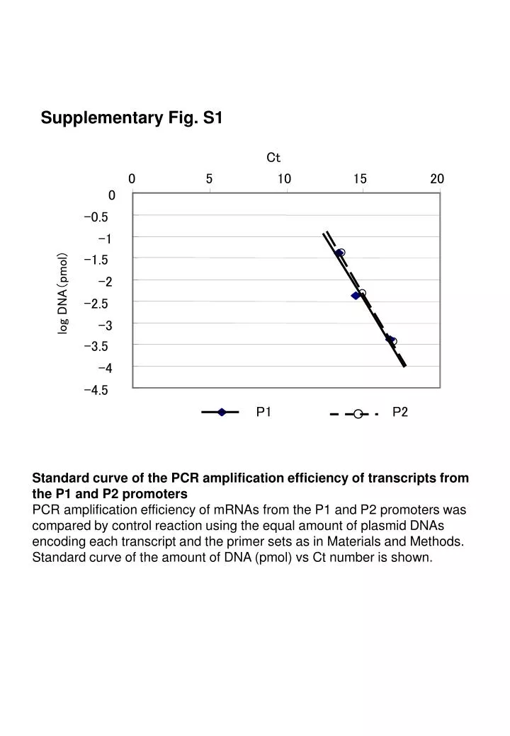
Supplementary Fig. S1 Ct 0 5 10 15 20 0 -0.5 -1 ○ -1.5 -2 ○ log DNA(pmol) -2.5 -3 ○ -3.5 -4 -4.5 P1 P2 ○ Standard curve of the PCR amplification efficiency of transcripts from the P1 and P2 promoters PCR amplification efficiency of mRNAs from the P1 and P2 promoters was compared by control reaction using the equal amount of plasmid DNAs encoding each transcript and the primer sets as in Materials and Methods. Standard curve of the amount of DNA (pmol) vs Ct number is shown.
Supplementary Fig. S2 Serum MMS -(h) - 0 1 2 3 4 5 6 9 0 1 2 3 4 5 6 9 ATF3 GAPDH 0 1 3 6 12 0 1 3 6 12(h) ATF3 b-tubulin 30 120 serum mRNA MMS mRNA 25 100 serum protein MMS protein 20 80 relative protein expression relative mRNA expression 15 60 10 40 5 20 0 0 0 6 12 h Differential response of the P1 and P2 promoters of human ATF3 gene to serum and genotoxic agent MMS HCT116 cells were serum-starved in the presence of 0.5 % serum for 48 h, and then stimulated by 20 % serum, or 100 mg / ml MMS. At each time indicated, ATF3 mRNA (□, ■) or protein (○, ●) was measured as in Methods. Data are means of three independent experiments with standard error bars.
Supplementary Fig. S3 A hATF3 GGCGGAGGTGGGGTTAGCTTCAGTTGACCAACCATGCCTTGAGGATAAATTGGATGGGAT mATF3 GGCAGGGGAGGGGTCAGCCTCAGACAACCAATCCTGCCTTGAGGATAAATTGTGTAGGAC *** * ** ***** *** **** ***** * ****************** * *** hATF3 CAGATGGGAAGATGTGACAAGAAGAGAAATCCTCCTCTATATAGGATGCTCTGCTGTTTC mATF3 CAGACCAGACAAGAGTATG-GAAGAGAGA--CTCCTCTGAACAGGATCTCCCACAGGGTC **** ** * * **** **** ******* * ***** * * * * hATF3 CTAAGGATTTTCAGCACCTTGCCCCAAAATG mATF3 CCAAGGAGTTCCAGCACTTCATCCCAAAATG * ***** ** *** *** * ********* B hATF3 ATTACGTCAGCCTGGGACTGGCAACACGGAGT-AAACGACCGCGCCGCCAGCCTGAGGGC mATF3 -----------CTGGGATTGGTAACCTGGAGTTAAGCGGGCTCCCTGCCAACGCGAGGGC ****** *** *** ***** ** ** * * * **** * ****** hATF3 TATAAAAGGGGTGATGCAACGCTCTCCAAGCCACAGTCGCACGCAGCCAGGCGCGCACTG mATF3 TTTAAAAGGGGTGATGCAACGCGCTCCCAGCCACAGTCTCACTCAGCGAGACGC-CGC-G * ******************** **** ********** *** **** ** *** * * * hATF3 CACAGCTCTCTTCTCTCGCCGCCGCCCGAGCGCACCCTTCAGCCCGCGCGCCGGCCGTGA mATF3 CACGGTGCTTCCC---------CAGTGGAGCCAATCGGCTAACCCGCGCTCCGGCA--GA *** * ** * * **** * * * ******* ***** ** hATF3 GTCCTCGGTGCTCGCCCGCCGGCCAGACAAACAGCCCGCCC---GACCCCGTCCCGACCC mATF3 GTCCTTGGCGCTCGCCCGCCGGCGGGACAGACCACCCGCCTCTGGCCGCTCTCTGGACCC ***** ** ************** **** ** ****** * * * ** ***** hATF3 TGGCCGCCCCGAGCGGAGCCTGGAGCAAAATG mATF3 TGGCCGCCCCGAGCGAAGACTGGAGCAAAATG *************** ** ************* Sequence alignment of the 5’ UTR of transcripts from the P1 and P2 promoters of human and mouse Sequence homology of the 5’ UTR of P1 transcripts of human (148 base; GC 47.3 %) and mouse (145 base; 53,1 %) is shown in A, and that of P2 transcripts of human (265 base; GC 69.4 %) and mouse (245 base; GC 66.9%) is shown in B.
Supplementary Fig. S4 Pol II 0.14 ■ □L428 ◆ ◇ DAUDI ■ 0.12 0.10 % Input 0.08 ■ ■ 0.06 ■ ■ ■ 0.04 ■ 0.02 □ □ □ ◆ □ □ □ ■ ◆ ◆ ◆ ◆ ◆ ◇ ◆ ◇ ◇ ◇ ◇ ◆ □ ◇ □ ◇ 0 -5 P1 +10 +20 +30 P2 +50 +55 kb LeoI (PAF1) 0.02 0.016 ■ ■ 0.012 % Input ■ ■ 0.008 ■ ■ ■ 0.004 ◆ ◆ ◆ □ ◆ ◆ □ □ ◇ ◆ ■ □ ◇ ◆ ◇ ◇ ◇ □ ◇ ◇ □ □ ◇ □ ◆ 0 -5 P1 +10 +20 +30 P2 +50 +55 kb Recruitment of RNA polymerase II onto the ATF3 gene in cancer cells L428 (■, □) or DAUDI (◆, ◇) cells were assayed for ChIP using anti-RNA polymerase II (upper panel) or anti-Leo1 antibody (lower panel) as in Methods. Immunoprecipitated DNA was measured by quantitative PCR throughout the human ATF3 gene and expressed as per cent of input DNA. Data represent the means of three independent experiments with standard error bars for control IgG (open) and specific antibodies (closed), respectively.
Supplementary Fig. S5 Panacetyl H3 0.7 ■ □L428 ◆ ◇ DAUDI ■ 0.6 0.5 % Input 0.4 0.3 0.2 ■ ■ ■ 0.1 ■ □ ■ ■ □ □ ■ □ □ □ □ □ 0 -5 P1 +10 +20 +30 P2 +50 +55 kb H3K4me3 1.0 ■ ■ 0.8 % Input 0.6 ■ ■ 0.4 ■ ■ ■ 0.2 ■ □ □ ◆ ◆ □ □ □ □ ◆ ◆ ◆ ◆ □ ◇ ◇ ◆ ◆ ◇ ◇ ◇ □ ◇ ◇ ◇ 0 -5 P1 +10 +20 +30 P2 +50 +55 kb Chromatin modification of the ATF3 gene in Hodgkin RS cells Immunoprecipitated DNA by anti-panacetyl H3 or anti-trimethyl H3K4 antibodies of L428 (■, □) or DAUDI(◆, ◇) cells was measured by quantitative PCR throughout the human ATF3 gene and expressed as per cent of the input. Data represent the means of three independent experiments with standard error bars for control IgG (open) and specific antibodies (closed), respectively.