Download
1 / 24
250 likes | 667 Views
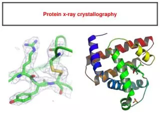
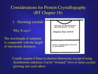
Protein Crystallography. At SSRL By: Silpa Nalam and Andrea Kurtz Advisors: Aina Cohen, Paul Ellis, and Nick Sauter. Project Description Protein Crystallization Web Page Design. Part 1: Introduction. Advantages of SSRL x-ray beam lines over home x-ray tubes higher intensity

E N D
Protein Crystallography At SSRL By: Silpa Nalam and Andrea Kurtz Advisors: Aina Cohen, Paul Ellis, and Nick Sauter
Project Description Protein Crystallization Web Page Design Part 1: Introduction
Advantages of SSRL x-ray beam lines over home x-ray tubes higher intensity tuning the x-rays allows one to choose one wavelength better resolution How is a diffraction pattern made? The pattern is made from diffracted x-rays by the atoms of the sample math and computers are used for interpretation because x-rays cannot be focused by a lens system dimensions of spots give unit cell information and their intensity gives the arrangement of the atoms in the unit cell Background on X-rays Diffraction Pattern
What is a Protein? Basic Structure polypeptide chain and its average size is 10,000 Daltons R groups have 20 different combinations and help the protein bind to ligands, catalyze chemical reactions and direct the polypeptide in its conformation Functions enzymes, structural elements, antibodies, hormones, and electron carriers
Protein Structure • 4 Levels of structure • give the protein a 3-D shape which helps carry out its functions • primary=sequence of R groups • secondary=random coil, alpha, or beta structures • tertiary=folding of secondary structures (biologically active) • quaternary=more than one polypeptide chain • Protein sensitivity • denaturation (protein unfolding) caused by heat, chemicals, changes in pH Alpha Helix Beta Sheet
Common Proteins in the Body Myoglobin is used as an oxygen storage molecule that can be found in the muscle tissue of vertebrates Hemoglobin is a unique blood protein that carries oxygen from the lungs to the rest of the body. Leghemoglobin is a type of hemoglobin found in plants. It is an important part of the nitrate fixation process
Crystallization with X-rays • Usefulness of x-rays • wavelength of x-rays is 10-8 cm • Difficulties in Crystallization • not usually in a crystalline fashion • not suited to be in crystalline form • protein has an extreme sensitivity • Why Crystallization? • provides the answer to many structure related questions
Background on Crystals • A crystal is a periodic arrangement of unit cells in a lattice. The unit cell is repeated in a 3-D lattice to make a crystal. • Common example of a crystal in a lattice is sodium chloride
Materials for Experiment Linbro Plate 24 well tray and each well is 1.5 cm in width and 1.8 cm in depth Myglobin The protein is placed in a reagent that reduces its solubility to close to precipitation. The concentration should be 5-50 mg/mL Cryo Solutions Allows for the crystals to grow. They can be bought or homemade. Gel The gel is used to ensure the coverslips stay over the well
Procedure For Experiment Vapor-Diffusion Method • The protein/precipitant mixture in the drop is less concentrated than in the well solution. • Water evaporates from the drop until the concentration of the precipitant in the drop is the same as in the well. • The conditions in the drop determine whether the protein will crystallize or not. Such as: ionic strength, temperature, and protein concentration. Finished Tray
Variables • As the precipitant point of the protein is approached, factors such as pH, temperature, ionic strength, and choice of buffer control whether the protein will separate from the solution as a crystal or a precipitate. • The pH, type of cryoprotectant (PEG or MPD) and buffer should be varied to get the protein in crystalline fashion. • It is hard to crystallize proteins under normal conditions because the crystal might react with water vapor. The crystals are usually grown in a cold room.
Key Points • Reproducibility • Crystallography: art or science? • Software based trials • Crystallization process • growing crystals • analysis of crystals • production of data • conclusion • theory of crystallization
Protein Crystallography at SSRL Part II: Determining Crystal Quality and Data Collection and Analysis
Background on Crystals • A body that is formed by the solidification of a chemical element, a compound, or a mixture and has a regularly repeating internal arrangement of its atoms and often external plane • faces • 7 Classes of Unit Cells • Described by three axes (a,b,c) and three angles (,,) Left: Single molecule of bovine pancreatic trypsin inhibitor (BPTI), considered the asymmetric unit in the unit cell below Below: Packing diagram for orthorhombic unit cell of BPTI in the most common space group, P212121 • 230 Space Groups • Characterized by symmetry elements such as rotational and screw axes • The presence of symmetry elements simplifies data collection and analysis.
Macromolecular Crystallization Crystallization of the protein is only the first step…
Examination of Samples Protein remains soluble Amorphous precipitate Gel precipitate Crystalline precipitate Cyclophilin crystals
Crystal Quality at a Glance Characteristics of Crystals Suitable for Analysis: • Large size, i.e., average length of approximately 200m • Single crystals with smooth, distinct faces and edges • Color (in many cases) or opacity (for myoglobin) Common Protein Crystal Defects: • Bunching or twinning • Size deficiencies • Denaturation with age
Mounting and Flash-Cooling Many protein crystals are extremely sensitive to changes in temperature and surrounding conditions (i.e., presence of original solvent). The crystal must be flash-cooled to 100K in a stream of liquid nitrogen to prevent damage from high-intensity x-rays. Teasing the crystal from the coverslip droplet Crystals grown in the cold room must be flash-cooled and transported to the experimental hutch with cryo-tools. Copper pin on goniometer head in liquid nitrogen stream
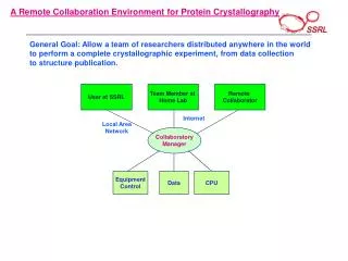
Data Collection Variables According to the size of the crystal and the type of protein, the following factors are assigned prior to data collection: • X-ray beam wavelength (on tuneable beam lines) • Crystal-to-detector distance • = degree of oscillation around phi axis • Exposure time • Start and end -angle Sample Control Panel at Beam Line 9-1
The Diffraction Pattern • Key Features: • spot separation • unit cell dimensions • spot intensity • unit cell contents Myoglobin diffraction pattern at 1.598Å resolution Left: Bragg’s Law and reflection of x-ray beams from crystal planes
Data Interpretation Using Mosflm, we can perform the following functions on the data embedded in the diffraction pattern: • Autoindexing and Cell Refinement • unit cell determination • mosaicity • orientation matrix • Integration • intensity: count-integrated and profile-fitted on each image • combine reflections on multiple images • Tests of Data Quality: • R-factor • Percentage of data measured • Systematic absences • I/I and F/F • Scaling and Merging • combining equivalent reflections • crystal breakdown and beam decay The importance of resolution in structural determination
Methods of Solving Structures • Direct - small molecules (less than 100 amino acids) with very high resolution data • Multiple Isomorphous Replacement (MIR) • Multiwavelength Anomalous Dispersion (MAD) • Molecular Replacement Method (MRM) Random Amplitudes Random Phases Above: Fourier synthesis diagrams. The red lines represent the model that was used to find the actual amplitudes and phases. Both factors are essential, since phases are not directly measurable and must be calculated from amplitudes
Future Improvements • Automated Batch Handling and X-Ray Diffraction Analysis • Integration of Computer Technology with Human Ability • Increased Internet-Based Interfacing for Data Collection, Analysis, Storage, and Retrieval Crystallographer’s Creed This is my x-ray machine. There are many like it, but this one is mine. My x-ray machine is my best friend. It is my life. I must master it as I must master my life. Without me, my x-ray machine is useless. Without my x-ray machine, I am useless. I must collect my data true. I must phase faster than my enemy who is trying to publish before me. I must shake him before he bakes me. I will. -Dr. Bernhard Rupp, LLNL