Download
1 / 70
881 likes | 1.52k Views
Neck Mass. Yonatan Avraham Demma. DIAGNOSIS. Patient history Physical examination Differential diagnosis Imaging studies Blood test. PATIENT HISTORY. The most important element in the evaluation of a neck mass is…. …the age of the patient.

E N D
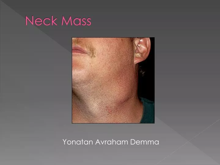
Neck Mass Yonatan Avraham Demma
DIAGNOSIS • Patient history • Physical examination • Differential diagnosis • Imaging studies • Blood test
PATIENT HISTORY The most important element in the evaluation of a neck mass is….
Most pediatric neck masses are inflammatory or congenital and resolve spontaneously or after appropriate medical therapy. In contrast, a neck mass in an adult over the age of 40 should be considered neoplastic in origin unless proven otherwise, particularly in the setting of tobacco or alcohol use.
PATIENT HISTORY • duration, • growth pattern, • and absence or presence of pain. • change in voice, • hoarseness, • difficulty with swallowing, • ear pain • generalized complaints: fever, night sweats, and weight loss. • patient's social history: alcohol and drug use, smoking, and recent travel.
PHYSICAL EXAMINATION • systematic investigation of all mucosal and submucosal areas of the head and neck. • mobility, consistency, and tenderness of the mass. • location of the neck mass is particularly important • Children: different branchial cyst • adult patients: virchov metastasis
ANATOMY Differential diagnosis
Anterior triangle bordered by anterior border of SCM, midline of neck, and mandible • muscular triangle formed by midline, superior belly of omohyoid, and SCM • carotid triangle formed by superior belly of omohyoid, SCM, and posterior belly of digastric • submental triangle formed by anterior belly of digastric, hyoid, and midline • submandibular triangle formed by mandible, posterior belly of digastric, and anterior belly of digastric
Posterior/Lateral triangle bordered by posterior border of SCM, trapezius, and clavicle • supraclavicular triangle formed by inferior belly of omohyoid, clavicle, and SCM • occipital triangle- formed by inferior belly of omohyoid, trapezius, and SCM
ANTERIOR NECK The structures that make up the anterior neck include • the larynx, • trachea, • esophagus, • thyroid and parathyroid glands, • carotid sheath, • and suprahyoid and infrahyoid strap muscles.
POSTERIOR/LATERAL NECK Contains: • lymph node, • the spinal accessory nerve, • the cervical plexus. • the brachial plexus • subclavianvessels.
Differential Diagnosis • Congenital neck mass • Inflammatory Neck mass: • Infectious • Non infectious • Neoplastic Disorder
CONGENITAL NECK MASSES • BRANCHIAL CLEFT CYSTS • THYROGLOSSAL DUCT CYSTS • LARYNGOCELES • PLUNGING RANULAS • LYMPHANGIOMAS • HEMANGIOMAS • TERATOMAS - DERMOID CYSTS • THYMIC CYSTS • STERNOCLEIDOMASTOID TUMORS OF INFANCY
1. BRANCHIAL CLEFT CYSTS • failure of the pharyngobranchial ducts to obliterate during fetal development. • most frequently present in late childhood or early adulthood when the cysts become infected usually after an upper respiratory tract infection. • tender, inflammatory mass located at the anterior border of the sternocleidomastoid muscle.
Classification three categories: • first, less than 1% • second, the most common • and third branchial cleft anomalies.
Treatment • initial control of the infection • surgical excision of the cyst and tract. incision and drainage procedures should be avoided
2. THYROGLOSSAL DUCT CYSTS • one-third of all congenital neck masses. • midline masses of the anterior neck . • may be asymptomatic and appear only when they become infected. • Thyroglossal duct cysts that occur off the midline may be difficult to differentiate from branchial cleft cysts. • pathognomonic sign on physical examination is vertical motion of the mass with swallowing and tongue protrusion, demonstrating the intimate relation to the hyoid bone.
TheSistrunk operation • excised with a cuff of tissue, including the center portion of the hyoid bone. • care is taken not to injure the hypoglossal nerves; • thyroid carcinomas can be present in a small percentage of thyroglossal duct cysts
3. LARYNGOCELES • abnormal dilation or herniation of the saccule of the larynx. • Laryngopyoceleis aSecondary infection of a laryngocele. • present with hoarseness, cough, dyspnea, dysphagia, a foreign body sensation. • Dx: Laryngoscopy, CT. • If symptomatic, Tx: • laryngoscopicdecompression for small lesions, • surgical excision through an external approach for larger lesions,
PLUNGING RANULAS • mucoceles of the floor of mouth • usually present as slow-growing, painless, submental masses. • arise from the sublingual gland and are defined as plunging when they extend through the mylohyoid muscle into the neck. • Tx: excision.
LYMPHANGIOMAS • congenital malformations of the lymphatic channels. • They arise owing to failure of the lymph spaces to connect to the remaining lymphatic system. • The mass is usually soft, doughy, smooth, nontender, and compressible. • can be transilluminated. • CT scanning and MRI are important studies both to delineate the extent of the disease • Tx: Surgical Debulkingbecause of the infiltrative nature
HEMANGIOMAS • malformations of vascular tissue. • usually present in the first few months of life, grow rapidly during the first year, and then begin to slowly involute at 18–24 months of age. • In 90% involution occurs without the need for any therapy. • present as a red or bluish soft mass that is compressible and increases in size with straining or crying. • Bruits may sometimes be auscultated over the lesion. • Dx: CT scans, MRI. • In the following symptoms: airway compromise, skin ulceration, dysphagia, thrombocytopenia, cardiac failure Systemic corticosteroids or surgical laser excision may be warranted in such cases.
TERATOMAS - DERMOID CYST approximately 3.5% of all teratomas. Their origin is from pluripotential cells, contain elements from all three germ layers. Usually present as midline, nontender, mobile neck masses and are most commonly noted at birth or within the first year of life. There is a 20% associated incidence of maternal polyhydramnios. can cause respiratory compromise or dysphagia secondary to compression. Dx: CT, MRI. Tx: Surgical excision.
THYMIC CYSTS • The third branchial pouch gives rise to the thymus during the 6th week of fetal life, elongates in the pharynx, and then descends into the mediastinum. • Thymiccysts arise when there is implantation of this thymic tissue along this descent. • present as slow-growing, asymptomatic masses that may be painful if infected. • On rare occasions, they grow rapidly and cause dyspnea or dysphagia. • CT scanning and MRI are useful in the differential diagnosis. • definitive diagnosis is made histologically by the presence of Hassall corpuscles. • Tx: surgical excision.
STERNOCLEIDOMASTOID TUMORS OF INFANCY • characterized histologically by dense fibrous tissue and the absence of normal striated muscle. • intimately related to congenital torticollis. • typically present as firm, painless, discrete masses within the sternocleidomastoid muscle; • slowly increase in size for 2–3 months and then regress for 4–8 months. • 80% resolve spontaneously and do not need any intervention other than physical therapy to prevent restrictive torticollis. • Surgical resection is reserved for persistent cases.
INFLAMMATORY NECK MASSES • Infectious inflammatory disorder: • Viral (reactive, HIV) • Bacterial (suppurative, toxoplasmosis, tularemia, brucellosis) • Granulomatous (cat-scratch disease, actinomycosis, atypical mycobacteria, tuberculosis, atypical tuberculosis, sarcoidosis) • Non infectious inflamatory disorder: • Sinus histiocytosis – Roni-Dorfman disease • Kawasaki disease • Castleman disease
VIRAL LYMPHADENOPATHY • REACTIVE VIRAL LYMPHADENOPATHY • HIV-ASSOCIATED INFLAMMATORY DISORDERS
REACTIVE VIRAL LYMPHADENOPATHY • the most common cause of cervical adenopathy in children. • usually associated with symptoms of an underlying upper respiratory tract infection. • The most common viral agents include adenovirus, rhinovirus, and enterovirus. • tend to regress in 1–2 weeks.
Management • Usually observation • a neck mass larger than 1 cm should be considered abnormal and warrant further investigation if it remains for more than 4–6 weeks or increases in size. • If persists, biopsies can be taken to search for other causes.
Mononucleosis • EBV can also present with lymphadenopathy • usually accompanied by the enlargement of other lymphoid tissues such as the adenoids or tonsils. • symptoms of fever and pharyngitis. • 4–6 weeks. • Tx: limited to supportive management.
HIV-ASSOCIATED INFLAMMATORY DISORDERS • Cervical Adenopathy or Persistent Generalized Lymphadenopathy • present in 12–45% of patients with human immunodeficiency virus (HIV). • other infectious or neoplastic etiologies must be ruled out • Tx: HIV Tx
BACTERIAL LYMPHADENOPATHY • Suppurative Lymphadenopathy • Toxoplasmosis • Tularemia • Brucellosis
Suppurative Lymphadenopathy • most frequently caused byStaphylococcus aureusand group AB-Streptococcus. • usually develop in the submandibular region • often accompanied by sore throat, skin lesions, and upper respiratory tract infection. • Empirical antibiotic therapy against anaerobic and gram-positive organisms is recommended as the first line of management. • If this fails, either FNA or incision and drainage may be indicated.
Toxoplasmosis • caused byToxoplasma gondii • contracted through the consumption of poorly cooked meat or the ingestion of oocytes excreted in cat feces. • present with fever, malaise, sore throat, and myalgias. • Dx: serologic testing. • Tx: ABx ex: sulfonamides (resprim).
Tularemia • caused byFrancisella tularensis • transmitted by rabbits, ticks, or contaminated water. • present with tonsillitis and painful adenopathy with systemic symptoms of fever, chills, headache, and fatigue. • Dx: Serology and Bc. • Tx: Streptomycin is the antibiotic of choice.
Brucellosis • Cause by Brucella. • most commonly transmitted to children by the ingestion of unpasteurized milk. • present with total body lymphadenopathy, fever, fatigue, and malaise. • Dx: Serology and BC. • Tx: Abx : trimethoprim–sulfamethoxazole or tetracycline
GRANULOMATOUS DISEASES • cat-scratch disease, • actinomycosis, • atypical mycobacteria, • tuberculosis, • atypical tuberculosis, • sarcoidosis.
Cat-Scratch Disease • Bartonellahenselae. • history of contact with cats can be elicited in 90% of cases. • more commonly seen in patients younger than 20 years. • tender lymphadenopathy, fever, and malaise. • lymphadenopathy typically preauricular and submandibular. • Dx: serologic testing. • generally benign and self-limited.
Actinomycosis • gram-positive bacillus. • 50% to 96% of cases of actinomycosis affect the head and neck regions. • painless, fluctuant, neck mass in the submandibular regions. • DX: histologically by the presence of granulomas with sulfur granules. • TX: Penicillin
Atypical Mycobacteria • typically presents in the pediatric population • unilateral neck mass located in the anterior triangle of the neck or parotid region. • brawny skin, induration, and pain. • Dx: culture and skin testing. • Tx: Surgical excision offers definitive treatment, although incision and curettage along with antibiotic therapy constitute an alternative management strategy.
Tuberculosis • more commonly in adults than in children. • Mycobacterium Tuberculosis • The presenting lymphadenopathy tends to be more diffuse and bilateral in contrast to atypical mycobacteria. • Tuberculin skin tests are strongly positive. • Cervical tuberculosis is also known asscrofula • Tx: antituberculous medications.
Sarcoidosis • presents most commonly in the second decade of life • lymph node enlargement, fatigue, and weight loss. • Chest radiography shows hilaradenopathy. • An elevated angiotensin-converting enzyme (ACE) level is seen in 60–90% of patients with sarcoidosis. • Dx: histologically by the presence of noncaseating granulomas. • TX: Corticosteroids.