Download
1 / 28
310 likes | 585 Views
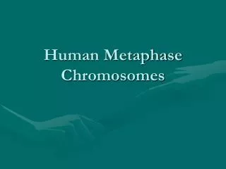
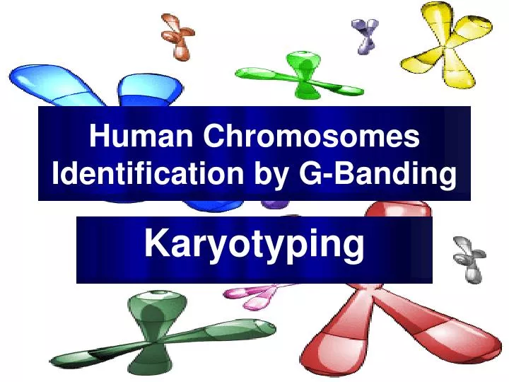
Human Chromosomes Identification by G-Banding. Karyotyping. Experiment Objectives. Preparing, Staining and Observing G-banding human chromosomes Develop an understanding of karyotyping and the association of various chromosomal abnormalities to diseases. Human Chromosomes.

E N D
Human Chromosomes Identification by G-Banding Karyotyping Mazen Zaharna Molecular Biology 1/2009
Experiment Objectives • Preparing, Staining and Observing G-banding human chromosomes • Develop an understanding of karyotyping and the association of various chromosomal abnormalities to diseases. Mazen Zaharna Molecular Biology 1/2009
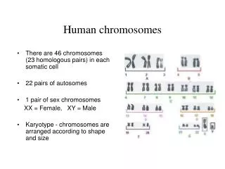
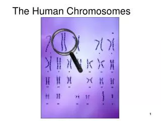
Human Chromosomes • A “normal” human carries 23 PAIRS of chromosomes (1 set came from the mother, 1 set came from the father) • 22 of these sets are called autosomes (or “self chromosomes”) • 1 set are the sex chromosomes • A female carries two X chromosomes (XX) • A male carries an X chromosome and a Y chromosome (XY) Mazen Zaharna Molecular Biology 1/2009
Why do scientists look at chromosomes? • Scientists can diagnose or predict genetic disorders by looking at chromosomes. • This kind of analysis is used in prenatal testing and in diagnosing certain disorders, such as • Down syndrome, • or in diagnosing a specific types of leukemia. Mazen Zaharna Molecular Biology 1/2009
Chromosome abnormalities • Chromosome abnormalities can be • numerical, as in the presence of • extra • or missing chromosomes, • or structural as in translocations, inversions, large scale deletions or duplications. Mazen Zaharna Molecular Biology 1/2009
Situations where analysis is strongly recommended • Problems with early growth & development • Fertility problems • Neoplasia • Pregnancy in older women Mazen Zaharna Molecular Biology 1/2009
What is a Karyotype? • A display or photomicrograph of an individual’s somatic-cell metaphase chromosomes that are arranged in a standard sequence (usually based on number, size, and type) Mazen Zaharna Molecular Biology 1/2009
Performing a Karyotype • The slides are scanned for metaphase spreads and usually 10 to 30 cells are analyzed under the microscope by a cytogeneticist. • When a good spread (minimum number of overlapping chromosomes) is found, a photograph is taken or the analysis is done by a computer. • The chromosomes are arranged in a standard presentation format of longest to shortest. Mazen Zaharna Molecular Biology 1/2009
How Do Scientists Identify Chromosomes? • Three key features to identify their similarities and differences: • Size. This is the easiest way to tell two different chromosomes apart. • Banding pattern. The size and location of Giemsa bands on chromosomes make each chromosome pair unique. • Centromere position. Centromeres are regions in chromosomes that appear as a constriction. • Using these key features, scientists match up the 23 pairs Mazen Zaharna Molecular Biology 1/2009
In metacentric chromosomes, the centromere lies near the center of the chromosome.Submetacentric & very Submetacentric chromosomes, have a centromere that is off-center, so that one chromosome arm is longer than the other. In acrocentric chromosomes, the centromere resides very near one end. Mazen Zaharna Molecular Biology 1/2009
Chromosome banding • Chromosomes are stained with various dyes enabling the chromosome segments to be identified • Most methods can distinguish 550 bands/ haploid set • High resolution methods can distinguish up to 850 bands/ haploid set that can allow identification of small interstitial deletions Mazen Zaharna Molecular Biology 1/2009
G-Banding Dye gives chromosomes a striped appearance because it stains the regions of DNA that are rich in adenine (A) and thymine (T) base pairs. Mazen Zaharna Molecular Biology 1/2009
G-Banding • Regions that stain as dark G bands replicate late in S phase of the cell cycle and contain more condensed chromatin, • While light G bands generally replicate early in S phase, and have less condensed chromatin. Mazen Zaharna Molecular Biology 1/2009
Chromosome Groups Mazen Zaharna Molecular Biology 1/2009
Chromosomal Abnormalities • Alterations in chromosome number. • Euploid - normal set (2n) • Polyploidy – extra set of the entire genome. • (3n, 4n etc) • Aneuploidy – the number of chromosomes is not a multiple of the normal haploid number. • Monosomy • one member of a chromosome pair is missing, (2n-1) • Trisomy • one chromosome set consists of 3 copies of a chromosome, (2n+1) Mazen Zaharna Molecular Biology 1/2009
Chromosomal abnormalities that can be detected by karyotyping Mazen Zaharna Molecular Biology 1/2009
Chromosomal abnormalities that can be detected by karyotyping Philadelphia Chromosome - CML Mazen Zaharna Molecular Biology 1/2009
Overview of Procedure • Collection of blood • Cell culture • Stopping the cell division at Metaphase • Hypotonic treatment of red & white blood cells • Fixation • Slide preparation Mazen Zaharna Molecular Biology 1/2009
Overview of Procedure • Slide dehydration • Treatment with enzyme • Staining Mazen Zaharna Molecular Biology 1/2009
Monitor the quality of chromosome spreading • Monitor the quality of chromosome spreading under phase contrast. • Chromosomes should be well spread • without visible cytoplasm, • should appear dark grey under phase contrast Mazen Zaharna Molecular Biology 1/2009
7-Slide dehydration • Place fixed, dry slides on slide rack in 60oC oven • Bake for 3 days • Allow to cool before proceeding to the next step Mazen Zaharna Molecular Biology 1/2009
8- Treatment with enzyme • Prepare 0.025% trypsin solution fresh, by mixing 5 ml of 0.25% trypsin with 45 ml Hank’s solution • Immerse slide in 0.025 % trypsin for 10-120 seconds • Remove slide from trypsin and immediately immerse in phosphate buffer to stop trypsin action Mazen Zaharna Molecular Biology 1/2009
Determination of Trypsin and Staining time Mazen Zaharna Molecular Biology 1/2009
9- Staining • Prepare a dilution of Giemsa stain by mixing 1 part of Giemsa stain with 3 parts of Phosphate buffer • Flood slide with Giemsa stain for 2 minutes • Rinse slides thoroughly with distilled water • Allow slides to drain, then place on 60oC slide warming tray until completely dry Mazen Zaharna Molecular Biology 1/2009
21 22 x y Mazen Zaharna Molecular Biology 1/2009