Download
1 / 21
210 likes | 445 Views
Knee and Hip Pain Quiz. Case 1. 20 yr old footballer. Swollen knee occurred after being tackled. Felt a popping sensation. Then instability on that leg + swelling. Knee gives way. Lachmann’s test positive. Anterior Cruciate Ligament Rupture (b).

E N D
Case 1 • 20 yr old footballer. • Swollen knee occurred after being tackled. • Felt a popping sensation. • Then instability on that leg + swelling. • Knee gives way. • Lachmann’s test positive.
Anterior Cruciate Ligament Rupture (b) • Lachmann's test determines anterior cruciate integrity. • The knee is flexed at 20-30 degrees and the leg is stabilized by the examiner sitting on the foot. The hamstrings should be relaxed. The leg and thigh are grasped firmly and moved in opposite directions. • Antero-posterior displacement of the tibia on the femur with the knee at 20-30 degrees and the muscles relaxed is a positive result.
Case 2 • 45 year old jogger. • Pain along medial knee joint line. • Mild swelling of knee, esp. after exercise. • Sensation of knee ‘locking’. • McMurray’s test positive.
Meniscal Tear (h) • McMurray’s = rotation test for demonstrating a torn meniscus. • Held by one hand which is placed along the joint line, the knee is flexed to 90ø while the foot is held by the sole with the other hand. The ankle is internally rotated and the knee extended. The manoeuvre is repeated with external rotation of the ankle and at varying degrees of knee flexion. • A tag, caused by a tear will cause a palpable or even audible click on extension of the knee. • The 'normal' leg must be checked for completeness: clicks can arise from normal tendon movement.
Case 3 • 15 yr old boy. • Knee ache, worse after activity. • Mild effusion. • Feeling of locking, where he has to flick the knee out to straighten it, especially after sitting for a long time. • X-ray shows white lesion, 0.2cm, in the knee joint.
Osteochondritis Dissecans (a) • Small segment of articular cartilage and subjacent bone may separate - dissect - as an avascular segment. • Caused by repeated minor trauma that produces an osteochondral fracture of a convex joint surface. • Characteristically affects young adult men. • Presents with intermittent pain, swelling and joint effusion of the affected joint. • The necrotic fragment may become detached and result in the joint 'locking' or 'giving way'. • Treatment is dependent upon the position of the fragment, but may include surgical removal.
Case 4 • 32 year old keen walker. • Pain just below patella. • Worse when walking, jumping and going down stairs.
Patella Tendinitis (e) • Inflammation of the patella tendon initiated usually by a small tear. • Commonly occurs in sports players. • Settles with rest and NSAIDS. A steroid injection around (not into) the tendon may also help
Case 5 • 80 year old female RH resident • Normally mobile • OA both knees • Sudden onset immobility and agitation • Lt knee rigid, swollen, hot, unable to move, extremely tender • Pyrexial & tachycardic
Septic arthritis (m) • Most common children, elderly • Predisposing factors: • Pre-existing joint disease (OA, RA) • Joint replacement • Injury or infection to affected leg • Bacterial infection affecting joint space • Requires admission for Abx, drainage. Can be very unwell
Case 6 • 13 yr old boy. • Keen sports player. • Pain + tenderness over tibial tuberosity. • Worse after playing sports.
Osgood Schlatter’s Disease (l) • Overuse syndrome associated with physical exertion before skeletal maturity. • It is a traction apophysitis caused by repeated avulsion of the apophysis into which part of the patellar tendon is inserted. • It usually occurs around the pubertal growth spurt when the quadriceps has enlarged but the apophysis has not yet fused to the tibia. • Usually resolves with a total embargo on sport for up to 6 months with gradual return afterwards
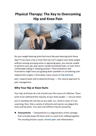
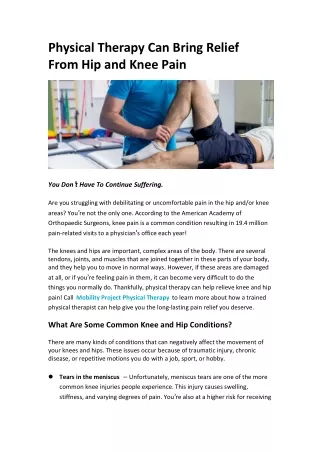
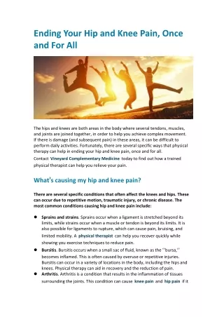
Case 7 • 55 year old woman • Obese for much of life • Increasing pain and stiffness in left hip • Also has pain in left knee • Now struggling to walk long distances • Rest relieves pain but joint is often stiff after prolonged rest
Osteoarthritis (o) • Single most common cause of locomotor disability • RFs include age, genetic predisposition, obesity + abnormal joint loading • Treatment involves analgesia, non-loading exercise, counselling. • Eventually prosthesis may be only option
Case 8 • 76 yr old man. • Acutely swollen, painful knee. • X-ray shows calcification of articular cartilage. • Joint aspirate contains bi-refringent crystals.
Pseudogout (k) • Due to deposition of calcium pyrophosphate dihydrate (CPPD) crystals in large joints, most commonly the knee. • The crystals are initially deposited in the cartilage - chondrocalcinosis - where they are associated with degenerative changes. • The shedding of the crystals into the joint space results in an acute synovitis and a clinical picture that is similar to that seen in gout. • In gout crystals are negatively bi-refringent.
Case 9 • 5 year old boy • Limping on right leg last month • C/o pain in right groin • Hip exam appears normal
Perthe’s disease (n) • Rare condition mainly affecting boys 3-10 years old • May be associated with trauma/infection • Bone death with reduced vascularisation, followed by remodelling of joint • Treatment included immobilisation, avoiding weight-bearing and surgery • Complications include early arthritis and premature fusion of growth plate
Case 10 • 32 year old male • Manual worker • 1 month of severe pain in right leg • Seems to ‘shoot’ down leg • Straight leg raise replicated this pain at 60 degrees
Sciatica (q) • Often mistaken for hip or even knee pathology • Low back pain is a major cause of long term disability • Three part management plan: • Analgesia to get moving • Exercise to build muscle strength • Physio to stretch back and teach good posture