Download
1 / 2
20 likes | 22 Views
Computed tomography (CT) is the current standard imaging modality for RTP because it provides electron density information that is fundamental for planning algorithms. However, CT is not always the best modality to identify the target volume because it is limited to discriminate between the tumour and adjacent soft tissues. Magnetic Resonance (MR) provides several advantages over CT, including high quality detailed images, excellent softtissue contrast, and the ability to distinguish between post-treatment fibrosis and tumour recurrence. On the other hand, CT attenuation maps are not available in MR and geometric distortion could be manifested due to the static magnetic field non-uniformities.

E N D
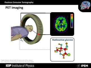
EDITORIAL The role of PET imaging in radiotherapy planning Alessandro Stefano PhD Stefano A. The role of PET imaging in radiotherapy planning. Nucl Med Radiol J. 2017;1(1)2-3. A The concept of planning target volumes (PTV) has been proposed by the International Commission on Radiation Units and Measurements (1) and it has been accepted and used in RTP. depending on the method used to delineate PET regions. BTV segmentation is a critical task due to lack of consistency in cancer contours, low resolution and high level of noise in PET imaging. The choice of a standard algorithm for BTV contouring is a very challenging yet unresolved step. Manual contouring is still extensively used in clinical environment, but it is user dependent and is time-consuming, because dozens of slices have to be manually segmented. For these reasons, many PET image segmentation methods have been proposed including thresholding and region growing methods that are the most widely used due to their implementation simplicity but they lack in robustness on low contrast and heterogeneous tumour regions. Other algorithms, such as variational approaches based on gradient differences between healthy and cancer tissues are mathematically efficient but noise sensitive and subjected to numerical fluctuation. Graph-based methods obtain efficient segmentation by using foreground and background seeds to locate the different tissues in the PET image. The seed identification can be automated; nevertheless, normal structures (i.e., brain, heart, bladder, and kidneys) will be wrong detected as the target seeds, leading to a wrong guidance to the segmentation, as suggested by Stefano et al. (6). Learning methods, such as artificial neural network, and support vector machine are efficient but require high computational steps. In addition, the high heterogeneity of PET images makes stable feature recognition very difficult. A comprehensive review of segmentation algorithms in PET imaging is given by Zaidi et al. (7). key issue in radiotherapy treatment planning (RTP) is how to deliver the radiation dose to cancer regions reducing the dose to healthy tissues. Over the past few decades, advances in hardware technologies have resulted in high-precision techniques such as conformal and intensity-modulated radiotherapy (IMRT). These techniques utilize non-invasive imaging to plan the radiotherapy treatment improving the relationship between the dose required to achieve local control and the expected treatment- related complications. Nevertheless, hardware precision in the radiation dose delivery is still greater than the software precision in target volume delineation. For this reason, target volume definition is recognized to be one of the most significant geometric uncertainties in RTP. Computed tomography (CT) is the current standard imaging modality for RTP because it provides electron density information that is fundamental for planning algorithms. However, CT is not always the best modality to identify the target volume because it is limited to discriminate between the tumour and adjacent soft tissues. Magnetic Resonance (MR) provides several advantages over CT, including high quality detailed images, excellent soft- tissue contrast, and the ability to distinguish between post-treatment fibrosis and tumour recurrence. On the other hand, CT attenuation maps are not available in MR and geometric distortion could be manifested due to the static magnetic field non-uniformities. In summary, accurate BTV detection is still a critical issue considering its substantial impact in RTP; any inaccuracy may lead to inadequate tumour coverage resulting in loco-regional recurrence. Studies have shown significant changes in PTV on the basis of PET information in patients with head and neck cancer (8), lung cancer (9), brain cancer (2) and cervical cancer (10). Other studies whose aim is to investigate the clinical utility of PET imaging in RTP are mandatory to validate and support the use of this technique in routine clinical practice. The integration of Positron Emission Tomography (PET) datasets in RTP may add another layer of sophistication in PTV definition: metabolic volume could be used to provide additional and complementary information with respect to anatomical imaging useful for treating the cancer region more precisely and to reduce any uncertainty and inter- and intra-observer variability in defining target volumes (2). PET imaging has been shown to decrease PTV reducing toxicity with the same radiation dose or enabling radiation dose escalation with the same toxicity (3). The “Biological Tumor Volume” (BTV) separates the tumour according to its biological activity. Accurate BTV definition is essential for escalating the radiation dose without increasing normal tissue injury. In addition, PET imaging has an enormous potential to improve patient management providing an in vivo measurement of the cancer’s biological processes (4). Metabolic changes are often faster and more indicative of the treatment effects than anatomical changes, providing a more rapid method to detect the therapy response. For this reason, PET imaging is being increasingly used for quantitative assessment of therapy response and for clinical trials of novel cancer therapies. Maximum Standardized Uptake Value (SUV) has been the first parameter introduced for PET study evaluation. It provides the highest uptake value inside the cancer region. Nevertheless, this value doesn’t provide any information about the cancer extension. Other quantitative parameters have been introduced, such as the BTV and the tumour lesion glycolysis (TLG) (5). TLG is able to provide both volumetric and metabolic information. It is calculated performing the multiplication of the mean SUV with the BTV value. For this reason, the identification of accurate and operator independent BTV segmentation algorithms is also mandatory to obtain accurate and reproducible PET parameters. REFERENCES 1. Berthelsen K, Dobbs J, Kjellén E, et al. What’s new in target volume definition for radiologists in ICRU report 71? How can the ICRU volume definitions be integrated in clinical practice? Cancer Imaging. 2007;7(1):104-16. 2. Rundo L, Stefano A, Militello C, et al. A fully automatic approach for multimodal PET and MR image segmentation in gamma knife treatment planning. Comput Methods Programs Biomed. 2017;144:77-96. 3. Niyazi M, Landrock S, Elsner A, et al. Automated biological target volume delineation for radiotherapy treatment planning using FDG- PET/CT. Radiat Oncol. 2013;8(1):180. 4. Allegra E, Saita V, De Natale M, et al. Use of PET/CT to detect local and regional laryngeal cancer recurrence after surgery. Rep Med Imaging. 2017;10:31-6. 5. Larson S, Erdi Y, Akhurst T, et al. Tumor treatment response based on visual and quantitative changes in global tumor glycolysis using PET- FDG imaging. The visual response score and the change in total lesion glycolysis. Clin Positron Imaging. 1999;2(3):159-171,. However, due to the nature of PET technique, the BTV varies substantially Institute of Molecular Bioimaging and Physiology, National Research Council (IBFM-CNR), Cefalù (PA), Italy Correspondence: Alessandro Stefano, Institute of Molecular Bioimaging and Physiology, National Research Council (IBFM-CNR), Cefalù (PA), Italy. email: alessandro. stefano@ibfm.cnr.it Received: November 22, 2017; Accepted: November 23, 2017; Published: November 30, 2017 This open-access article is distributed under the terms of the Creative Commons Attribution Non-Commercial License (CC BY-NC) (http:// creativecommons.org/licenses/by-nc/4.0/), which permits reuse, distribution and reproduction of the article, provided that the original work is properly cited and the reuse is restricted to noncommercial purposes. For commercial reuse, contact reprints@pulsus.com OPEN ACCESS Nucl Med Radiol J Vol 1 No 1 November 2017 2
Stefano management of head and neck squamous cell carcinoma. Egypt J Radiol Nucl Med. 2011;42(2):157-67 6. Stefano A, Vitabile S, Russo G, et al. An enhanced random walk algorithm for delineation of head and neck cancers in PET studies. Med Biol Eng Comput. 2016;55(6):1-12. 9. Zheng Y, Sun X, Wang J, et al. FDG-PET/CT imaging for tumor staging and definition of tumor volumes in radiation treatment planning in non-small cell lung cancer. Oncol Lett. 2014;7(4):1015-20. 7. Zaidi H, El Naqa I. PET-guided delineation of radiation therapy treatment volumes: A survey of image segmentation techniques. Eur J Nucl Med Mol Imaging. 2010;37(11):2165-87. 10. Salem A, Salem A, Ibraheem A et al. Evidence for the use PET for radiation therapy planning in patients with cervical cancer: A systematic review. Hematol Oncol Stem Cell Ther. 2011;4(4):173-81. 8. Khodary M, Tabashy R, Omar W et al. The role of PET/CT in the Nucl Med Radiol J. Vol 1 No 1 November 2017 3