Download
1 / 4
40 likes | 48 Views
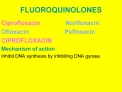
Ciprofloxacin is an antibiotic used in the treatment of various bacterial infections, such as bone and joint infections, respiratory tract infections, intra-abdominal infections, certain type of infectious diarrhea, typhoid fever, skin infections, and urinary tract infections, among others. For some<br>infections it is used besides other antibiotics. It can be taken orally or used intravenously.

E N D
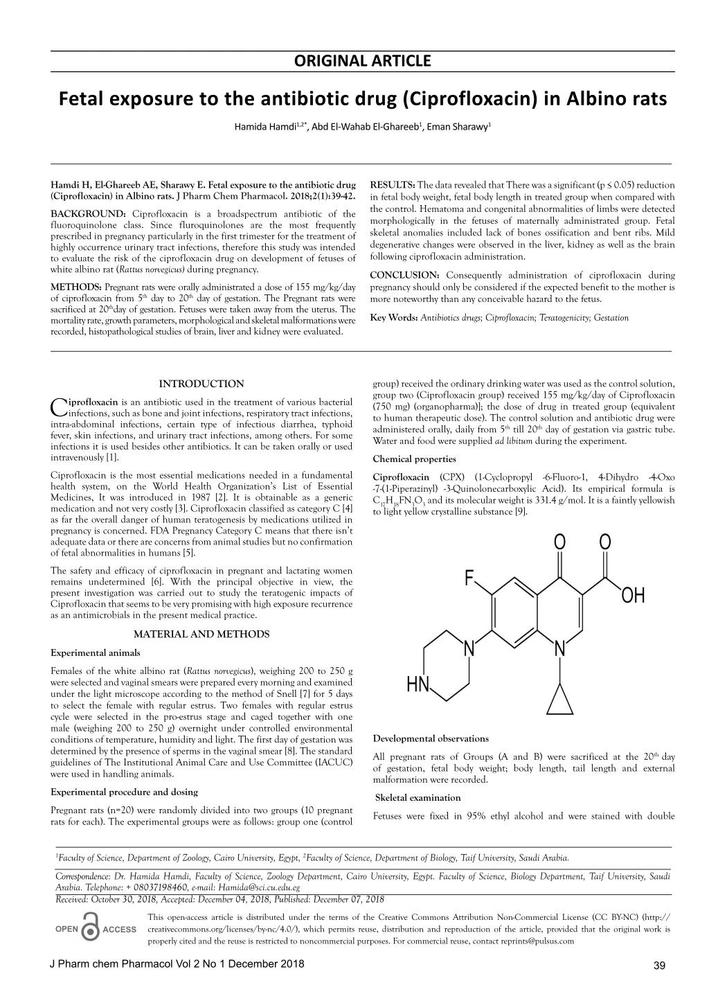
ORIGINAL ARTICLE Fetal exposure to the antibiotic drug (Ciprofloxacin) in Albino rats Hamida Hamdi1,2*, Abd El-Wahab El-Ghareeb1, Eman Sharawy1 Hamdi H, El-Ghareeb AE, Sharawy E. Fetal exposure to the antibiotic drug (Ciprofloxacin) in Albino rats. J Pharm Chem Pharmacol. 2018;2(1):39-42. RESULTS: The data revealed that There was a significant (p ≤ 0.05) reduction in fetal body weight, fetal body length in treated group when compared with the control. Hematoma and congenital abnormalities of limbs were detected morphologically in the fetuses of maternally administrated group. Fetal skeletal anomalies included lack of bones ossification and bent ribs. Mild degenerative changes were observed in the liver, kidney as well as the brain following ciprofloxacin administration. BACKGROUND: Ciprofloxacin is a broadspectrum antibiotic of the fluoroquinolone class. Since fluroquinolones are the most frequently prescribed in pregnancy particularly in the first trimester for the treatment of highly occurrence urinary tract infections, therefore this study was intended to evaluate the risk of the ciprofloxacin drug on development of fetuses of white albino rat (Rattus norvegicus) during pregnancy. CONCLUSION: Consequently administration of ciprofloxacin during pregnancy should only be considered if the expected benefit to the mother is more noteworthy than any conceivable hazard to the fetus. METHODS: Pregnant rats were orally administrated a dose of 155 mg/kg/day of ciprofloxacin from 5th day to 20th day of gestation. The Pregnant rats were sacrificed at 20thday of gestation. Fetuses were taken away from the uterus. The mortality rate, growth parameters, morphological and skeletal malformations were recorded, histopathological studies of brain, liver and kidney were evaluated. Key Words: Antibiotics drugs; Ciprofloxacin; Teratogenicity; Gestation group) received the ordinary drinking water was used as the control solution, group two (Ciprofloxacin group) received 155 mg/kg/day of Ciprofloxacin (750 mg) (organopharma)]; the dose of drug in treated group (equivalent to human therapeutic dose). The control solution and antibiotic drug were administered orally, daily from 5th till 20th day of gestation via gastric tube. Water and food were supplied ad libitum during the experiment. INTRODUCTION C intra-abdominal infections, certain type of infectious diarrhea, typhoid fever, skin infections, and urinary tract infections, among others. For some infections it is used besides other antibiotics. It can be taken orally or used intravenously [1]. iprofloxacin is an antibiotic used in the treatment of various bacterial infections, such as bone and joint infections, respiratory tract infections, Chemical properties Ciprofloxacin is the most essential medications needed in a fundamental health system, on the World Health Organization’s List of Essential Medicines, It was introduced in 1987 [2]. It is obtainable as a generic medication and not very costly [3]. Ciprofloxacin classified as category C [4] as far the overall danger of human teratogenesis by medications utilized in pregnancy is concerned. FDA Pregnancy Category C means that there isn’t adequate data or there are concerns from animal studies but no confirmation of fetal abnormalities in humans [5]. Ciprofloxacin (CPX) (1-Cyclopropyl -6-Fluoro-1, 4-Dihydro -4-Oxo -7-(1-Piperazinyl) -3-Quinolonecarboxylic Acid). Its empirical formula is C17H18FN3O3 and its molecular weight is 331.4 g/mol. It is a faintly yellowish to light yellow crystalline substance [9]. The safety and efficacy of ciprofloxacin in pregnant and lactating women remains undetermined [6]. With the principal objective in view, the present investigation was carried out to study the teratogenic impacts of Ciprofloxacin that seems to be very promising with high exposure recurrence as an antimicrobials in the present medical practice. MATERIAL AND METHODS Experimental animals Females of the white albino rat (Rattus norvegicus), weighing 200 to 250 g were selected and vaginal smears were prepared every morning and examined under the light microscope according to the method of Snell [7] for 5 days to select the female with regular estrus. Two females with regular estrus cycle were selected in the pro-estrus stage and caged together with one male (weighing 200 to 250 g) overnight under controlled environmental conditions of temperature, humidity and light. The first day of gestation was determined by the presence of sperms in the vaginal smear [8]. The standard guidelines of The Institutional Animal Care and Use Committee (IACUC) were used in handling animals. Developmental observations All pregnant rats of Groups (A and B) were sacrificed at the 20th day of gestation, fetal body weight; body length, tail length and external malformation were recorded. Experimental procedure and dosing Skeletal examination Pregnant rats (n=20) were randomly divided into two groups (10 pregnant rats for each). The experimental groups were as follows: group one (control Fetuses were fixed in 95% ethyl alcohol and were stained with double 1Faculty of Science, Department of Zoology, Cairo University, Egypt, 2Faculty of Science, Department of Biology, Taif University, Saudi Arabia. Correspondence: Dr. Hamida Hamdi, Faculty of Science, Zoology Department, Cairo University, Egypt. Faculty of Science, Biology Department, Taif University, Saudi Arabia. Telephone: + 08037198460, e-mail: Hamida@sci.cu.edu.eg Received: October 30, 2018, Accepted: December 04, 2018, Published: December 07, 2018 This open-access article is distributed under the terms of the Creative Commons Attribution Non-Commercial License (CC BY-NC) (http:// creativecommons.org/licenses/by-nc/4.0/), which permits reuse, distribution and reproduction of the article, provided that the original work is properly cited and the reuse is restricted to noncommercial purposes. For commercial reuse, contact reprints@pulsus.com OPEN ACCESS J Pharm chem Pharmacol Vol 2 No 1 December 2018 39
Hamdi et al. staining of fetal skeletons for cartilage (Alcian blue) and bone (Alizarin red) according to the method de-scribed by Neubert et al. [10]. Histopathological preparation Parts of fetal tissues (Liver, kidney, brain) of two groups were fixed in 10% neutral buffered formalin then stored in 70% alcohol for a general histological preparation. The fetal tissues were dehydrated in ascending grades of ethyl alcohol, cleared in terpineol and embedded in paraffin wax. Serial transverse sections 5 microns thick of different fetal tissues were cut, mounted and stained with haematoxylin and eosin for general histological studies [11]. Figure 1)Photographs of uterus of pregnant rat at the 20th day of gestation (A) Control uterus, showing symmetrical distribution of fetuses in the two uterine horns. V = Vagina. (B and C) Uterus of treated pregnant rats, (B) Showing partial resorption and asymmetrical distribution of fetuses, (C) Showing complete resorption STATISTICAL ANALYSIS All the values were presented as means (μ) ± standard errors of the means (S.E.M) comparison between more than two different groups was carried out using the one-way analysis of variance (ANOVA). RESULTS Effects of ciprofloxacin during the gestational period (from 5th to 20th day of gestation) I. Pregnant albino rats Morphological studies External symptoms and mortality: The pregnant rats orally administered with 155 mg/kg (Group B) of ciprofloxacin during the gestational period (5th -20th day). No external signs of toxicity, no mortality cases were recorded; all the treated dams were survived to the end of study. Figure 2) Photographs of fetuses at 20th day of gestation, (A): normal fetus, showing normal growth, (B): fetus of maternally treated with 155 mg/kg ciprofloxacin, showing growth retardation and excencephaly as well as transparence skin, (C): fetus of maternally treated showing hematoma at the hind limb (arrow), (D): fetus maternally treated showing hematoma at the back (arrow) Change in body weight gain: All over the gestational period, the maternal body weight was followed, for the control and experimental group. The average maternal body weight was recorded (Table 1). There was a reduction in maternal weight gain in the treated group (B) when compared with the control group (no significant difference across groups, p ≥ 0.05). TABLE 1 Showing effect of ciprofloxacin on fetal body weight, fetal body length, tail length, placental/weight and mother weight gain at 20th day of gestation. Average weight of placenta: The average weight of placenta of the treated pregnant rats group was decreased as compared to control (Table 1). There was no significant difference (p ≥ 0.05). Fetus length (F.L) Placenta weight (P.WT) Mother weight gain (M.WT) Fetus weight (F.WT) Tail length (T.L) Morphological observations of uterus: The uterus of control pregnant rats on day 20 of gestation showed normal distribution of the implanted fetuses in the two horns (Figure 1A). The uterus of treated pregnant rats showed partial resorption in only one horn, asymmetrical distribution of fetuses in the two uteri horns and reduced number of fetuses, uterine horns showing visible embryonic resorped sites (Figures 1B and 1C). Groups Control (A) 4.57 ± 0.12 5.98 ± 0.05 1.55 ± 0.05 0.51 ± 0.01 52.42 ± 8.43 155 mg/Kg Group (B) 3.15 ± 0.13a 2.08 ± 0.27a 1.29 ± 0.21 0.49 ± 0.05 41.19 ± 3.56 Effect of ciprofloxacin on fetuses II. Growth retardation proximal parts of the ilium and ischium of the pelvic girdle. Ribs of control fetuses, on the 18th day of gestation, acquired a normal set of 13 pairs of ribs; each of which consists of cartilaginous parts (the sternal portion of ribs) and a fully ossified part (the vertebral portion of ribs) as demonstrated in Figure 3A. The sternum of the control fetuses at 20th day of gestation consists of six well ossified pieces called sternebrae, of which the first one is large and called maubrium and the last one is also large and called xiphisternum. The last sternebra is terminated with an expanded cartilaginous plate called xiphoid cartilage. Each sternebrae is formed from two symmetrical bars. They are fused together the ventral mid-line. The cartilaginous junctions of the sternebrae are articulated with the sternal ribs or costal cartilages of the first seven pairs of ribs. On the other hand, fetuses of the maternally treated group with 155 mg/kg ciprofloxacin showed lack of ossification of roof of the skull, caudal vertebra and Phalanges, The rudimentary 13th ribs (Figure 3B). Absence of ossification of metacarpals, metatarsals, caudal vertebra, phalanges, curved 12th rib and the rudimentary 13th ribs (Figure 3C). The morphological examination of the fetuses showed that the ciprofloxacin caused growth retardation represented by a decrease in fetal body weight, body length and tail length (Table 1). There was a significant (p ≤ 0.05) reduction in fetal body weight, fetal body length in treated group when compared with the control group (A). Morphological malformations The fetus from control pregnant rats appeared with normal shape, correct weight and length (Figure 2A). The gross pathology of fetuses per dam was represented in Figures 2B-2D. The most observed anomalies were, hematoma (red patches at different parts of bodies), fore and hind limbs malformations, exencephaly, and short snout. Skeletal examination On the 20th day of gestation, the cleared cartilage and bone preparations of control rat fetuses have designated that in all parts of the axial skeleton skull, vertebrae and ribs as well as appendicular skeleton comprising the fore and hind limbs, pectoral and pelvic girdles, both chondrification and ossification processes have been obviously completed as demonstrated in Figure 3A, displaying the developed cartilage and bone in the different parts of skeleton of these fetuses. The cartilaginous parts of the skull included the proximal part of the nasal, the tympanic, squamosal and supra occipital regions. In the vertebral column, the proximal ends of the transverse processes and vertebral arches have cartilaginous ends. Also, two thirds of the caudal vertebrae and sternal portions of the ribs appeared cartilaginous. In addition to the cartilaginous joints in between the long bones of both fore and hind limbs, cartilaginous parts were found in the distal end of the scapula as well as Histopathological studies Examination of serial transverse sections of the brain, liver and kidney of albino rat fetuses maternally treated with 155 mg/kg ciprofloxacin on the 20th day of gestation showed some histopathological changes. Liver of fetuses The histological examination of the hepatic tissue of fetuses of the control group showed normal histological structure (Figure 4A). The hepatic tissue of fetuses maternally treated with 155 mg/kg ciprofloxacin revealed some histological changes such as golden yellow pigment localized extra and intracellular with multiple lymphoid cells, megakaryoblasts and fatty J Pharm chem Pharmacol Vol 2 No 1 December 2018 40
Fetal exposure to the antibiotic drug (Ciprofloxacin) in Albino rats 155 mg/kg ciprofloxacin revealed some histological changes such as multi focal areas of renal tubular necrosis, degenerated glomeruli and oedema in between the tubules and glomeruli was detected (Figures 5B and 5C). Brain The brain tissues of fetuses from control pregnant rats showed, normal features under microscopic observation as in cerebrum, while in fetuses maternally treated with 155 mg/kg ciprofloxacin showing vacuolization in the matrix of striatum in the cerebrum and dark neurons (Figures 6A and 6B). No any pathological changes detected in cerebellum. Figure 3) Photographs of skeleton of fetuses at 20th day of gestation, (A): Skeleton of control fetus showing well ossification (B): Skeleton of fetus maternally treated with 155 mg/kg ciprofloxacin, showing lack of ossification of skull (long arrow), rudimentary rib (short arrow). (C): Showing incomplete 12th and 13th ribs (rudimentary ribs) (black arrow), lack ossification of central vertebra (arrow), non- ossification of .caudal vertebra (circle) and no ossification of Tarsus DISCUSSION Wisely use of medications is gradually becoming the focus of fervency in medical practice. Recently, the interest has manifested by repeated, both preclinical and clinical, testing of new medications or medicinal agents all over the world. Although the toxic ability of drugs to produce congenital anomalies in offsprings when given to pregnant mothers did not gain much importance before the well-known disaster caused by Thalidomide, it is now well recognized that both extensive preclinical and post-marketing monitoring are main requirements to avoid parallel disaster in future. All medications that have been established as teratogens in man can be shown to be teratogenic in animals. Furthermore, medications shown to be teratogenic in animals may be teratogenic in man under proper conditions of dosage and timing, this fact was borne out by the value of animal screening [12]. The current work revealed that Ciprofloxacin treatment induced fetal resorption, and a significant reduction in the weight, length of fetuses, these results are also in accordance with Siddiqui and Naqvi [13]. We suggested that the growth retardation of embryos due to degeneration of the trophoblast and decidual cell and incomplete formation of the placenta, which play an important role in the transmission of nutrients to the embryo as reported in the study of Kurebe et al. [14], also, these results may be attributed to deficiency of nutritional supply from mother to fetuses due the diarrhea caused to female rats receiving ciprofloxacin or which might be attributed to imbalance in intestinal microflora as reported in the study of Takayama et al. and Watanabe et al. [15,16]. Figure 4) Photomicrographs of fetal Liver section at 20th day of gestation, (A) a control fetal section, showed normal histological structure hepatocytes (H) and central vein (CV). (H&E, 200x) (B): Section of liver of fetus maternally treated with 155 mg/kg ciprofloxacin, showing fatty degeneration (FD) in hepatocytes, pigmentation (blue arrow), and leucocytic cells permeation (black arrow), (C): Section of liver of fetus maternally treated showing golden yellow pigment (~) localized extra and intracellular with multiple lymphoid cells and megakaryoblasts (~) in between the hepatocytes (H). H&E 40x These findings are in agreement with the findings of many other researchers [4,17,18], who reported the teratogenic effects of ciprofloxacin in women exposed to it during pregnancy. Congestion, hematoma, fore and hind limb defects, short snout, exencephaly were the most repeated anomalies observed. In addition, a broad variety of incompletely ossified, unossified skull bones (mostly nasal, frontal, parietal and interparietal), abnormal vertebrae, appearance of supernumerary ribs, incompletely ossified, unossified bones of fore and hind limbs and absence of ossification of metacarpals, metatarsals and phalanges. The skeletal changes recorded in our study are similar to those described previously by others [19-22], these results are also in agreement with the findings of Lemus [23] who reported that severe alterations in the development of embryo cartilage and bones were associated with enrofloxacin and ciprofloxacin administration, These results agreed with that recorded by many researchers, following ofloxacin administration to pregnant rats and rabbits [15]; levofloxacin administration to rats [16]; Norfloxacin administration to pregnant rats [24]. Stahlmann [25] noted that fluroquinolone induced delay in the developmental phase of the epiphyseal growth with growth inhibition. The bone and cartilage damage could be due to fluoride accumulation with repeated fluroquinolone administration reported by Arora [26]. Fluroquinolones caused defect in cartilage development and delay the ossification process. A wide spectrum of musculoskeletal complications that involve not only tendon but also cartilage, bone, and muscle are associated with Fluoroquinolone antibiotics [27]. Figure 5) Photomicrographs of fetal kidney section at the 20th day of gestation, (A): Section of kidney of control fetal, showed normal histological structure of glomeruli (G) and proximal tubules (X). H&E X 100 (B): Section of kidney of fetus maternally treated, showing multi focal areas of renal tubular necrosis (arrows) and degenerated glomeruli (DG). (H&D, 200x), (C): Section of kidney of fetus maternally treated, showing odema in between the tubules and glomeruli and marked degeneration of the epithelial cells lining the renal tubules (arrow). H&E 100x… In the present study, the liver of fetuses maternally treated with 155 mg/kg ciprofloxacin showed golden yellow pigment localized extra and intracellular with multiple lymphoid cells, megakaryoblasts and fatty degeneration in between the hepatocytes. Figure 6) Photomicrographs of fetal brain section at the 20th day of gestation, (A): Section of control fetal brain, showing normal histological structure of the cerebrum. H&E 100x, (B): Section of brain of fetus maternally treated with 155 mg/kg ciprofloxacin, showing vacuolization (arrows), dark neuron (~). H&E 100x In this experiment, the kidney of fetuses maternally treated group revealed some histopathological changes such as multi focal areas of renal tubular necrosis, degenerated glomeruli and oedema in between the tubules and glomeruli was detected. These findings are in agreement with the study of Mohamed et al. [24], who reported that Norfloxacin administration induced hypoplasia or atrophy of one or both kidneys, also, Ofloxacin administration to rats and rabbits [28]. degeneration in between the hepatocytes (Figures 4B and 4C). Kidney of fetuses The kidney of fetus of control group showed normal histological structure (Figure 5A). Meanwhile sections of kidney of fetuses maternally treated with J Pharm chem Pharmacol Vol 2 No 1 December 2018 41
Hamdi et al. 16. Watanabe, Fujikawa T, Harada KS, et al. Reproductive toxicity of the new quinolone antibacterial agent levofloxacin in rats and rabbits. Arzneimittel-Forschung A. 1992;42:374–7. In this experiment, the histopathological changes in the fetal brain of the treated groups were evident in the early stages of development. As already noted, vacuolization in the matrix of striatum in the cerebrum and dark neurons. Which might ciprofloxacin which easily cross blood brain barrier and compete with gamma-aminobutyric acid receptor as reported by Akahane et al. [29]. 17. Friedman JM, Polifka JE. Teratogenic effects of drugs: A resource for clinicians (TERIS). 2nd edn. 2000. Baltimore: Johns Hopkins University Press, USA. These results could be attributed to the inhibitory effect of fluoroquinolones on DNA gyrase, the enzyme essential for negative super helical twisting into double stranded DNA (a reaction that generates tension in the double helix that favors unwinding of double strands) and the catenation and decatenation of two duplex DNA circles interlocked like links in a chain [30,31]. On the other hand DNA damage induced by CPX may be attributed to its ability to releasing oxygen free radicals [32]. It is known that mutations caused by Oxygen free radicals attack DNA [33]. 18. Schaefer C, Amoura Elefant E, Vial T, et al. Eur J Obstet Gynecol Reprod Biol. 1996;69-83. 19. Kim JG, Yun HI, Shin HC, et al. Embryo lethality and teratogenicity of a new fluoroquinolones antibacterial DW-116 in rats. Arch Toxical. 2000;74:120-4. 20. Kim JG, DH, Shin SH, et al. Developmental toxicity evaluation of the new fluoroquinolones antibacterial DW-116 in rats. Teratogenesis Carcinogenesis Mutagenesis. 2003;1:123-36. CONCLUSION 21. Kim JG, Shin DH, Kim SH, et al. Peri-and postnatal developmental toxicity of the fluoroquinolone antibacterial DW-116 in rats. Food Chem Toxicol. 2004;42:389-95. It can be concluded that thus administration of ciprofloxacin during pregnancy should only be considered if the expected benefit to the mother is greater than any possible risk to the fetus. 22. Kim JG, Shin DH, Kim SH, et al. Developmental toxicity assessment of the new flouruquinolone antibacterial DW-116 in rabbits. J Appl Toxicol. 2005;25:52-9. REFERENCES 1. The American Society of Health System Pharmacists. Ciprofloxacin Hydrochloride. 2015. 23. Lemus JA, Blanco G, Arroyo B, et al. Fatal embryo chondral damage associated with fluoroquinolones in eggs of threatened avian scavengers. Environ Pollut. 2009;157:2421-7. 2. World Health Organization, WHO.19thedn. Model List of Essential Medicines (PDF), 2015. 24. Mohamed A, Mohamed E, Ahmed S, et al. Embryotoxic and Teratogenic Effects of Norfloxacin in Pregnant Female Albino Rats. Advances in Pharmacological Sciences. 2014;6:1-6. 3. Hamilton, Richard J. Tarascon pharmacopoeia. 15th edn. Jones & Bartlett Publishers. 2014; 85. 4. Nahum GG, Uhl K, Kennedy DL. Antibiotic use in pregnancy and lactation: what is and is not known about teratogenic and toxic risks. Obstet Gynecol. 2006;107:1120-38. 25. Stahlmann R. Children as a special population at risk-quinolones as an example for xenobiotics exhibiting skeletal toxicity. Archives of Toxicology. 2003;77:7–11. 5. Greenfield M. Commonly used antibiotics in pregnancy. 2004. 26. Arora NK. Are fluoroquinolones safe in children? Indian Journal of Pediatrics. 1994;61:601–3. 6. Net Doctor. NetDoctor: the UK’s leading independent health website. 7. Snell GD. Biology of the laboratory Mouse. The Blakiston Company, Philadeliphia, USA. 1956. 27. Hall MM, Finnoff JT, Smith J. Musculoskeletal complications of fluoroquinolones: Guidelines and precautions for usage in the athletic population, Journal of Injury, Function and Rehabilitation. 2011;3:132–42. 8. McClain RM, Becker BA. Teratogenicity, foetal toxicity and placental transfer of lead nitrate in rats. Toxicol Appl Pharmacol. 1975;931:72-82. 28. Davis GJ, McKenzie BE. Toxicologic evaluation ofofloxacin, American Journal of Medicine C. 1989;87:43S–6S. 9. US Food and Drug Administration. Cipro Labeling Revision Supplement 073 (PDF), 2009. 29. Akahane K, Kato M, Takayama S. Involvement of inhibitory and excitatory neurotransmitters in levofloxacin-and ciprofloxacin-induced convulsions in mice, Antimicrobial Agents and Chemotherapy. 1993;37:1764–70. 10. Neubert D, Merkerd HJ, Kwasigroch P. Double staining of foetal skeletons for cartilage and bone in methods in prenatal toxicology. 1977. 11. Selim MJ, Afm SI. A study on the teratogenic effect of ciprofloxacin. Bangladesh J Physiol Pharmacol. 2006;22:9-11. 30. Wolfson KS, Hooper DC. Fluoroquinolone antimicrobial agents, Clinical Microbiology Reviews. 1989;2:378–424. 12. Banchroft JD, Stevens A, Turner DR. Theory and practice of Hitological Techniques. 4th edn; Churchil Living Stone, New York-London-San Francisco-Tokyo, 1996. 31. Vancutsem PM, Babish JG, Schwark WS. The fluoroquinolone antimicrobials: structure, antimicrobial activity, pharmacokinetics, clinical use in domestic animals and toxicity. The Cornell Veterinarian. 1990; 80:173–86, 13. Siddiqui MA, Naqvi SNH. Evaluation of the teratogenic potentials of ciprofloxacin in albino rat. J Morphol Sci. 2010;27:14-8. 32. Gürbay A, Gonthier B, Signorini-Allibe N, et al. Ciprofloxacin-induced DNA damage in primary culture of rat astrocytes and protection by vitamin E. Neurotoxicol. 2006;27:6-10. 14. Kurebe M, Asaoka H, Moriguchi M. Toxicological studies on a new cephamycin, MT-141. IX. Its teratogenicity test in rats and rabbits, Japanese Journal of Antibiotics. 1984;37:1186–210. 33. Arriaga Alba M, Rivera Sanchez R, Parra Cervantes G, et al. Anti- mutagenesis of b-carotene to mutations induced by quinolone on Salmonella typhimurium. Arch Med Res. 2000;31:156–61. 15. Takayama S, Watanabe T, Akiyama Y. Reproductive toxicity of ofloxacin, Arzneimittel-Forschung. 1986;36: 1244–8. J Pharm chem Pharmacol Vol 2 No 1 December 2018 42