Download
1 / 1
10 likes | 104 Views
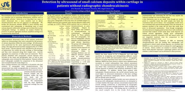
Detection by ultrasound of small calcium deposits within cartilage in patients without radiographic chondrocalcinosis Eric Russell, DO; Humaira Hussain, MD; Angel Checa, MD Drexel University College of Medicine and Hahneman University Hospital. Results. Results (cont.). Discussion (cont.).

E N D
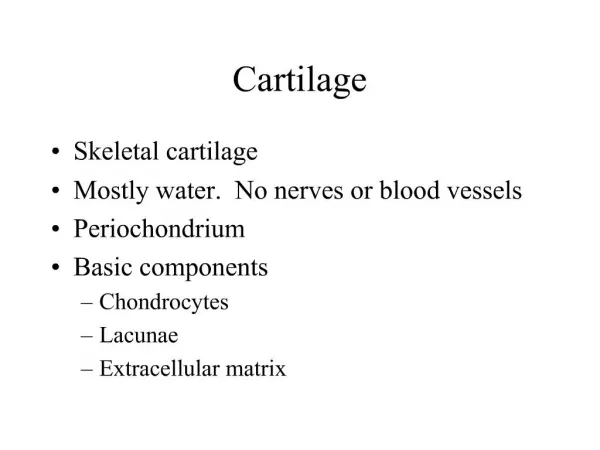
Detection by ultrasound of small calcium deposits within cartilage in patients without radiographic chondrocalcinosis Eric Russell, DO; Humaira Hussain, MD; Angel Checa, MD Drexel University College of Medicine and Hahneman University Hospital Results Results (cont.) Discussion (cont.) Introduction Of the 353 patients who underwent MSKUS, 24 (6.8%) patients had MSKUS evidence of aggregates of calcium within the wrist or knee cartilage. Two (8%) patients were excluded from the study since prior radiographs of the joint were not available (Figure 1). MSKUS detected more patients with calcium deposits within joint cartilage (p=<0.001). Fifteen (63%) patients had no evidence of LFC Musculoskeletal ultrasound (MSKUS) has increasingly been used as a sensitive tool in assessing inflammatory arthritis such as rheumatoid arthritis.1 Gutierrez et al. described two cases in which ultrasound was a more sensitive tool to detect chondrocalcinosis that was not apparent on radiograph.7 We further the discussion whether MSKUS is more sensitive than conventional joint radiography in identifying cases of small calcium deposits within joint cartilage and correlate this to prior radiographic findings. In addition, we analyze whether the presence of calcium aggregates facilitate amplified cartilage loss as measured by ultrasound. predominately had underlying OA and this may explain the reduced cartilage loss seen in either group. A few limitations exist in our study. Even though our results point to a possible trend, the small sample size limits the conclusions that can be drawn. Our sonographic results were not correlated with synovial fluid analysis since the majority of patients enrolled in the study did not have this study done prior to ultrasound examination. Even though sonographic chondrocalcinosis imaging patterns have been well established, synovial fluid analysis would verify these small deposits are indeed calcium aggregates. Finally, prior radiographic examination primarily consisted of AP and lateral views and not tangential (sunrise) view for comparison to the sonographic transverse view. Considering the low sensitivity of MRI in detecting chondrocalcinosis and our finding that radiography was insufficient to detect small calcium deposits, we conclude that MSKUS is a very sensitive tool to evaluate for small aggregates of calcium. Future studies are needed to evaluate whether these small deposits represent a pathologic pattern and also whether a clinical impact can be attributed to their presence MFC Tr A C Figure 2: Patient with sonographic but not radiographic chondrocalcinosis. (A) Non-magnified sonographic view of the left knee in transverse view revealing small calcium deposits within the medial compartment. (B) Magnified medial knee compartment. (C) Radiograph of the same knee without evidence of calcium deposition. . LFC=Lateral femoral condyle, Tr=trochlea, MFC=Medal femoral condyle. chondrocalcinosis on prior radiographs (Figure 2). Seven (29%) had evidence of chondrocalcinosis on conventional radiograph (Figure 3). Those patients that had sonographic, but had no radiographic calcium deposits, predominately had small calcium deposition. The group with radiographic chondrocalcinosis often showed calcium deposits too extensive to measure on ultrasound (Table 1). Cartilage thickness was not different between patients with or without radio-graphic chondrocalcinosis [mean (mm) = 1.71 vs. 1.61 respectively, p = 0.70 with unpaired t-test, Table 2]. B Materials & Methods Musculoskeletal ultrasound scans of 353 patients performed within our rheumatology clinic were evaluated for the presence of calcium deposition within the joint cartilage. All ultrasounds were performed by the same experienced ultrasonographer using a GE Logiq ultrasound machine equipped with a 8-13 MHz linear probe. Calcium deposits were identified by the presence of hyperechoic foci within the joint cartilage or fibrocartilage and they were measured along their longest axis. Calcium aggregates too numerous or bulky were not measured. Those patients identified with sonographic calcium deposits were included in the study and demographic data, as well as past radiographs, were reviewed for these patients. Patients without prior radiographs available for comparison to the ultrasound were excluded from the study (Figure 1). Knee cartilage thickness was evaluated in all patients who had evidence of sonographic knee chondrocalcinosis. This measurement was made sonographically on transverse view of the hyperflexed knee. Measurements from three separate comp- LFC MFC Tr A B Figure 3. Patient with sonographic and radiographic chondrocalcinosis. (A) Non-magnified sonograph of the left knee in transverse view showing extensive calcium deposition within the medial and lateral compartments of the knee (solid arrow). (B) Radiograph of the same knee showing bi-compartment chondrocalcinosis (arrow outline). LFC=Lateral femoral condyle, Tr=trochlea, MFC=Medal femoral condyle. Discussion MSKUS has been shown to be a diagnostic tool with high specificity in recognizing and distinguishing different crystal deposits such as CPPD and monosodium urate.2,5,6 Sonographic patterns of crystal deposition have been well described.2,3,4,5 In addition, MSKUS has been shown to detect more inflamed knees secondary to crystal arthropathies than clinical assessment alone.6 Our study demonstrated that musculoskeletal ultrasound was better than conventional radiography in detecting small calcium deposits in patients with symptoms attributed to other joint pathology. In addition, among those patients with radiographic chondrocalcinosis, the overall calcium burden was much higher on ultrasound examination of the knee cartilage suggesting conventional radiography detects calcium deposits that are already quite extensive. Because the sonographic calcium burden was more extensive in the group with radiographic chondrocalcinosis, we attempted to correlate whether this larger calcium concentration impacted the overall cartilage architecture as determined by sonographic measurement of the knee cartilage thickness. Knee cartilage loss was similar in both groups. This finding may be a reflection of the small sample size in the study. Both groups References 1. Szkudlarek, M., E. Narvestad, M. Klarlund, et al. 2004. Ultrasonography of the metatarsophalangeal joints in rheumatoid arthritis: Comparison with magnetic resonance imaging, conventional radiography, and clinical examination. Arthritis Rheum (2004) 50:2103– 2112. 2. Filippucci E., Gutierrez M., Georgescu D., Salaffi F., Grassi W. Hyalin cartilage involvement in patients with gout and calcium pyrophosphate deposition disease. An ultrasound study. Osteoarthritis and Cartilage (2009)17:178. 3. Frediani B., Filippou G., Falsetti P., Lorenzini S., Baldi F., Acciai C., Siagkri C., Marotto D., Galeazzi M., Marcolongo R.. Diagnosis of calcium pyrophosphate dihydrate crystal deposition disease: ultrasonographic criteria proposed. Ann Rheum Dis (2005) 64:638–640. 4. Ciapetti A., Filippucci E., Gutierrez M., Grassi W. Calcium pyrophosphate dihydrate crystal deposition disease: sonographic findings. Clin Rheumatol (2009) 28:271–276. 5. W. Grassi, MD,* G. Meenagh, MD,† E. Pascual, MD,‡ and E., Filippucci, MD “Crystal Clear”—Sonographic Assessment of Gout and Calcium Pyrophosphate Deposition Disease. Semin Arthritis Rheum. (2007) 37(2):133-4. 6. Filippucci E, Scirè CA, Delle Sedie A, Iagnocco A, Riente L, Meenagh G, Gutierrez M, Bombardieri S, Valesini G, Montecucco C, Grassi W. Ultrasound imaging for the rheumatologist. XXV. Sonographic assessment of the knee in patients with gout and calcium pyrophosphate deposition disease. Clin Exp Rheumatol. (2010) 28(1):2-5. 7. Gutierrez M., Di Geso L., Filipucci E., Grassi W. Calcium Pyrophosphate Crystals Detected by Ultrasound in Patients without Radiographic Evidence of Cartilage Calcifications. J Rheumatol (2010) 37:2602-2603. Patients underwent MSKUS (13 months) N=353 Chondrocalcinosis on MSKUS (N=24) artments (medial and lateral femoral condyle and the trochlea) within the knee were made and the calculated mean was deter-mined. The compartment with the most significant measured cartilage loss was noted. Patients with both sonographic chondrocalcinosis and radiograph for comparison (N=22) Excluded patients with sonographic chondrocalcinosis without radiographs for comparison (N=2) Acknowledgements Statistical analysis provided by Edward Gracely, PhD Associate Professor, Department of Epidemiology and Biostatistics Drexel University College of Medicine. Drs. Carolyn O’Connor and Vincent Zarro, Drexel University College of Medicine Figure 1: Flow chart of study selection process.