Download
1 / 36
400 likes | 660 Views
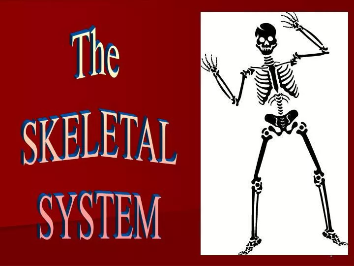
The SKELETAL SYSTEM. THE SKELETAL SYSTEM: OVERVIEW. I. INTRODUCTION The organs of the skeletal system include the bones and the structures that connect bones to other structures, including ligaments, tendons, and cartilages. The adult skeleton is composed of 206 separate bones.

E N D
The SKELETAL SYSTEM
THE SKELETAL SYSTEM: OVERVIEW I. INTRODUCTION • The organs of the skeletal system include the bones and the structures that connect bones to other structures, including ligaments, tendons, and cartilages. • The adult skeleton is composed of 206 separate bones.
Division of skeleton • Axial skeleton • Includes the bones of: • skull, vertebral column, and rib cage. • These bones are involved in protection, support, and carrying other body parts. • Appendicular skeleton • Bones of upper & lower limbs and the girdles (shoulder bones and hip bones) that attach them to the axial skeleton. • Involved in locomotion and manipulation of the environment.
AXIAL SKELETON Appendicular SKELETON
AXIAL SKELETON • 1) Skull: 28 irregular shaped bones from the skull. • The skull consists of two major division: • Cranium is formed by eight bones. • Face is formed by fourteen bones.
2) VERTEBRAL COLUMN Divided into 5 main regions Cervical spine (7) Thoracic spine (12) Lumbar spine (5) Sacrum (5) Coccyx (4) The 5 sacrum vertebrae and 4 coccyx vertebrae are fused to form one solid bone. AXIAL SKELETON
THORAX 12 pairs of ribs Joined to thoracic vertebrae Top 10 ribs joined to sternum Remaining two have “free” ends – ‘floating’ AXIAL SKELETON
Forms mainly the extremities of the body and their connections to the axial skeleton Consists of - limbs (arms & legs) - shoulder and pelvic girdles APPENDICULAR SKELETON
4 types of bones: Long Bones Much longer than they are wide. All bones of the limbs except for the patella (kneecap), and the bones of the wrist and ankle. Consists of a shaft plus 2 expanded ends. Short Bones Roughly cube shaped. Bones of the wrist and the ankle. Bone Classification Femur Carpal Bones
Types of bones: Flat Bones Thin, flattened, and usually a bit curved. Scapulae, sternum, (shoulder blades), ribs and most bones of the skull. Irregular Bones Have weird shapes that fit none of the 3 previous classes. Vertebrae, hip bones, 2 skull bones ( sphenoid and the ethmoid bones). Bone Classification Sternum Sphenoid Bone
Parts of long bone 1) diaphysis: main shaft like structure, its hollow cylindrical shape, its functions of providing strong support bone. 2) epiphysis: both end of long bone, epiphysis have a bulbous shape that provide generous space near joint. • For muscle attachment and give stability of joint.
Parts of long bone 3) articular cartilage thin layer of hyaline cartilage that covers joint surface. 4) Periosteum: dense white fibrous membrane that cover bone except at joint surface and anchoring bone to muscles. 5) Marrow cavity: a tubelike hollow space in the diaphysis of long bone. 6) endosteum: a thin epithelial membrane that line the medullry cavity.
Composition of bone matrix • Inorganic salts: the hardness of bone result from the deposition of high specialized chemical crystal of calcium and phosphorous called hydroxyapatite, the process called calcification in addition to mg, Na, sulphate and fluoride. • Organic substance: collagenous fiber and mixture protein and poly saccarids called ground substance, provide support and adhesion between cellular and fiberous. • Chondroitin and glucosamine are required for repair and maintenance of bone and cartilage.
Types of bone cells 1. Osteoblasts • Bone-forming cells found in all bone surfaces Bone-building cells. • Synthesize and secrete collagen fibers and other organic components of bone matrix. • serve as a framework for the deposition of calcium and phosphate calcification The blue arrows indicate the osteoblasts. The yellow arrows indicate the bone matrix they’ve just secreted.
Types of bone cells 2. Osteoclasts • Giant multinucleate cells • Responsible for the active erosion of bone minerals • Contain large numbers of mitochondria and lysosomes
Types of bone cells • 3. Osteocytes—mature, nondividing osteoblast surrounded by matrix, lying within lacunae
Bone Marrow • Specialized type of soft, diffuse connective tissue; called myeloid tissue • Site for the production of blood cells • Found in medullary cavities of long bones and in the spaces of spongy bone
Microscopic Structure of Compact Bone • Contains many cylinder-shaped structural units calledosteons, or Haversian systems • Four types of structures make up each osteon: • Lamella—concentric, cylinder-shaped layers of calcified matrix • Lacunae—small spaces containing tissue fluid in which bone cells are located between hard layers of the lamella
Microscopic Structure of Compact Bone • Canaliculi—ultrasmall canals radiating in all directions from the lacunae and connecting them to each other and to the Haversian canal • Haversian canal—extends lengthwise through the center of each osteon and contains blood vessels and lymphatic vessels
Bone Marrow • Two types of marrow are present during a person’s lifetime: • Red marrow • Found in virtually all bones in an infant’s or child’s body • Functions to produce red blood cells • Yellow marrow • As an individual ages, red marrow is replaced by yellow marrow • Marrow cells become saturated with fat and are no longer active in blood cell production
Bone Marrow • The main bones in an adult that still contain red marrow include the ribs, bodies of the vertebrae, the humerus, the pelvis, and the femur • Yellow marrow can alter to red marrow during times of decreased blood supply, such as with anemia, exposure to radiation, and certain diseases
Functions of Bone • Support—bones form the framework of the body and contribute to the shape, alignment, and positioning of the body parts • Protection—bony “boxes” protect the delicate structures they enclose • Movement—bones with their joints constitute levers that move as muscles contract • Mineral storage—bones are the major reservoir for calcium, phosphorus, and other minerals • Hematopoiesis—blood cell formation is carried out by myeloid tissue
Regulation of Blood Calcium Levels • Skeletal system (cont.) • Homeostasis of calcium ion concentration essential for the following: • Bone formation, remodeling, and repair • Blood clotting • Transmission of nerve impulses • Maintenance of skeletal and cardiac muscle contraction
Regulation of Blood Calcium Levels • Mechanisms of calcium homeostasis • Parathyroid hormone • Primary regulator of calcium homeostasis • Stimulates osteoclasts to initiate breakdown of bone matrix and increase blood calcium levels • Increases renal absorption of calcium from urine • Stimulates vitamin D synthesis
Mechanisms of calcium homeostasis (cont.) • Calcitonin • Protein hormone produced in the thyroid gland • Produced in response to high blood calcium levels • Stimulates bone deposition by osteoblasts • Inhibits osteoclast activity • Far less important in homeostasis of blood calcium levels than parathyroid hormone
Fracture Types • Open (compound) bone ends penetrate the skin. • Closed (simple) bone ends don’t penetrate the skin. • Spiral ragged break caused by excessive twisting forces. Sports injury/Injury of abuse • Greenstick bone breaks incompletely. One side bent, one side broken. Common in children whose bone contains more collagen and are less mineralized
Osteomalacia Literally “soft bones.” Includes many disorders in which osteoid is produced but inadequately mineralized. Causes can include insufficient dietary calcium Insufficient vitamin D fortification or insufficient exposure to sun light. Rickets Children's form of osteomalacia More detrimental due to the fact that their bones are still growing. Signs include bowed legs, and deformities of the pelvis, ribs, and skull. Clinical Conditions What about the above x-ray is indicative of rickets?
Osteomyelitis Inflammation of bone and bone marrow caused by pus-forming bacteria that enter the body via a wound (e.g., compound fracture) or migrate from a nearby infection. Fatal before the advent of antibiotics. Clinical Conditions
Clinical Conditions • Osteoporosis • Osteoporosis is a disease that thins and weakens the bones to the point that they become fragile and break easily. Women and men with osteoporosis most often break bones in the hip, spine, and wrist, but any bone can be affected. You can't "catch" osteoporosis or give it to someone else.
Any questions ??? The end