Download
1 / 52
520 likes | 641 Views
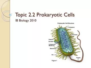
Characteristics of Cells and Life. Prokaryotic Cells. All living things (single and multicellular ) are made of cells that share some common characteristics: - basic shape – spherical, cubical, cylindrical - internal content – cytoplasm , surrounded by a membrane

E N D
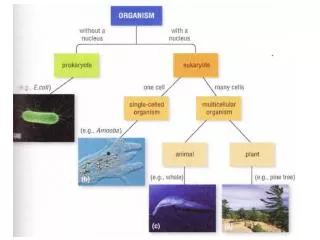
Characteristics of Cells and Life Prokaryotic Cells All living things (single and multicellular) are made of cells that share some common characteristics: • - basic shape – spherical, cubical, cylindrical • - internal content – cytoplasm, surrounded by a membrane • DNA chromosome(s), ribosomes, metabolic capabilities Two basic cell types: eukaryotic and prokaryotic
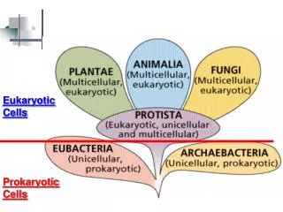
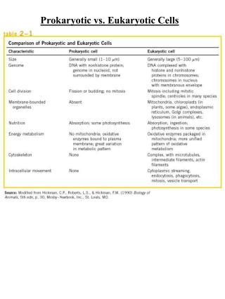
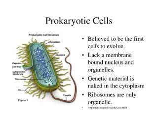
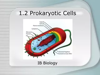
Characteristics of Cells Prokaryotic Cells Eucaryotic cells: animals, plants, fungi, and protists • contain double-membrane bound nucleuswith DNA • chromosomes • contain membrane-bound organellesthat • compartmentalize the cytoplasmand perform specific • functions Procaryotic cells: bacteria and archaea - no nucleus or other membrane-bound organelles
Characteristics of Life What is life ? Prokaryotic Cells • Growth and development • Reproduction and heredity – genome composed of DNA packed in • chromosomes; produce offspring sexually or asexually • Metabolism – chemical and physical life processes • Movement and/or irritability – respond to internal/external stimuli; • self-propulsion of many organisms • Cell support, protection, and storage mechanisms – cell walls, vacuoles, • granules and inclusions • Transport of nutrients and waste
Prokaryotic Profiles: Bacteria and Archaea Prokaryotic Cells
Prokaryotic Profiles: Bacteria and Archaea Prokaryotic Cells
External Structures Prokaryotic Cells - Appendages • two major groups of appendages: • Motility – flagella and axial filaments (periplasmic • flagella) • Attachment or channels – fimbriae and pili - Glycocalyx – surface coating
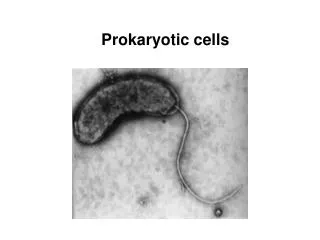
External Structures - Flagella 3 parts: • 1. filament – long, thin, helical structure composed of protein • flagellin • 2. hook- curved sheath • 3. basal body – stack of rings firmly anchored in cell wall Rotates 360o Number and arrangement of flagella varies: • monotrichous, lophotrichous, amphitrichous, peritrichous -> Functions in motility of cell through environment Prokaryotic Cells
External Structures - Flagella Prokaryotic Cells Gram-negative Gram-positive
External Structures – Flagella Arrangements Prokaryotic Cells Monotrichous Lophotrichous single flagellum at one end small bunches arising from one end of cell Amphitrichous Peritrichous flagella at both ends of cell flagella dispersed over surface of cell; slowest
External Structures – Operation of Flagella Signal sets flagella into rotary motion: Prokaryotic Cells • counterclockwise –> results in smooth linear direction –> run • clockwise -> cell stops and tumbles Guide bacteria in a direction in response to external stimulus: • -> chemical stimuli – chemotaxis; positive and negative • -> light stimuli – phototaxis
External Structures – Chemotaxis Prokaryotic Cells
External Structures – Axial Filaments Prokaryotic Cells Periplasmic, internal flagella, enclosed between cell wall and cell membrane of spirochetes Produce cellular motility by contracting and imparting twisting or flexing motion
External Structures – Other Appendages: Fimbriae Prokaryotic Cells Fine, proteinaceous, hairlike bristles from the cell surface Function in adhesion to other cells and surfaces -> do not provide locomotion -> pathogens use it to attach tightly to epithelial cells -> infection
External Structures – Other Appendages: Pili Prokaryotic Cells Rigid tubular structure made of pilin protein Found only in Gram negative cells Function to join bacterial cells for partial DNA transfer called conjugation -> sex pili
External Structures – Glycocalyx Coating of molecules external to the cell wall, -> made of sugars and/or proteins Two types: • -> slime layer - loosely organized and attached • -> capsule - highly organized, tightly attached Functions: • -> protect cells from dehydration and nutrient loss • -> inhibit killing by white blood cells by phagocytosis contributing to pathogenicity • -> attachment - formation of biofilms Prokaryotic Cells
External Structures – Glycocalyx -> Biofilms Prokaryotic Cells Staphylococcus aureus
The Cell Envelope -> External covering outside the cytoplasm -> Composed of two basic layers: • cell wall and cell membrane -> Maintains cell integrity -> Two generally different groups of bacteria demonstrated by Gram stain: • Gram-positive bacteria: thick cell wall composed primarily of peptidoglycan and cell membrane • Gram-negative bacteria: outer cell membrane, thin peptidoglycan layer, and cell membrane Prokaryotic Cells
The Cell Envelope Prokaryotic Cells
The Cell Envelope: Cell Wall -> Determines cell shape, prevents lysis (bursting) or collapsing due to changing osmotic pressures Peptidoglycan is primary component: • unique macromolecule composed of a repeating framework of long glycan chains cross-linked by short peptide fragments Prokaryotic Cells
Peptidoglycan Prokaryotic Cells
The Cell Envelope: Gram-Positive Cell Wall Prokaryotic Cells Thick, homogeneous sheath of peptidoglycan • -> 20-80 nm thick • -> includes teichoic acidand • lipoteichoic acid: function in cell • wall maintenance and enlargement • during cell division • -> move cations across the cell • envelope • -> stimulate a specific immune • response
The Cell Envelope: Gram-Negative Cell Wall -> Composed of an outer membrane and a thin peptidoglycan layer -> Protective structure while providing some flexibility and sensitivity to lysis -> Periplasmic space surrounds peptidoglycan -> Outer membrane is similar to cell membrane bilayer structure -> contains lipopolysaccharides and lipoproteins (LPS) -> function as receptors and block immune response -> contains porin proteins in upper layer – regulate molecules entering and leaving cell -> release exotoxins Prokaryotic Cells Porin (channel for small molecule).
The Cell Envelope: Comparison of Cell Walls Prokaryotic Cells
The Cell Envelope: Comparison of Gram + and Gram- Prokaryotic Cells
The Gram Stain -> Differential stain that distinguishes cells with a Gram-positive cell wall from those with a Gram-negative cell wall • Gram-positive - retain crystal violet and stain purple • Gram-negative - lose crystal violet and stain red from safranin • counterstain -> Important basis of bacterial classification and identification -> Practical aid in diagnosing infection and guiding drug treatment Prokaryotic Cells
The Gram Stain Prokaryotic Cells
Atypical Cell Walls Some bacterial groups lack typical cell wall structure i.e. Mycobacterium and Nocardia • Gram-positive cell wall structure with lipid mycolic acid (cord factor) -> pathogenicity and high degree of resistance to certain chemicals and dyes -> basis for acid-fast stain used for diagnosis of infections caused by these microorganisms Some have no cell wall i.e. Mycoplasma • -> cell wall is stabilized by sterols • -> pleomorphic (variations in shape) Prokaryotic Cells
Degradation of Cell Walls Lysozyme cleaves Peptidoglycan Penicillin inhibits enzyme responsible for construction of peptidoglycan • Gram-positive cell exposed to one of these agents • -> cell wall completely loosened -> protoplast • Gram-negative cell exposed to one of the agents • -> peptidoglycan loosened but outer membrane retained -> spheroplast Prokaryotic Cells
Cell Membrane Phospholipid bilayer with embedded proteins – fluid mosaic model Functions in: • -> providing site for energy reactions, nutrient processing, and • synthesis • -> transport into and out of the cell Prokaryotic Cells
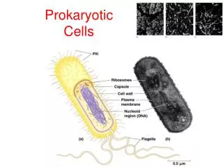
Bacterial Internal Structures Cell cytoplasm: • -> dense gelatinous solution of sugars, amino acids, and salts • -> 70-80% water • serves as solvent for materials used in all cell functions Prokaryotic Cells
Bacterial Internal Structures Chromosome • -> single, circular, double-stranded DNA molecule that • contains all the genetic information required by a cell • -> DNA is tightly coiled around a protein, aggregated in a • dense area called the nucleoid. Prokaryotic Cells
Bacterial Internal Structures Plasmids • -> small circular, double-stranded DNA • -> free or integrated into the chromosome • -> duplicated and passed on to offspring • -> not essential to bacterial growth and metabolism • -> may encode antibiotic resistance, tolerance to toxic metals, • enzymes and toxins • -> used in genetic engineering- readily manipulated and • transferred from cell to cell Prokaryotic Cells
Bacterial Internal Structures Ribosomes • -> made of 60% ribosomal RNA and 40% protein • -> consist of two subunits: large and small • -> procaryotic differ from eucaryotic ribosomes in size and • number of proteins • -> site of protein synthesis • -> present in all cells Prokaryotic Cells
Bacterial Internal Structures Inclusions and granules • -> intracellular storage bodies • -> vary in size, number and • content • -> Bacterial cell can use them • when environmental sources • are depleted. • examples: glycogen, poly-b-hydroxybutyrate, gas vesicles for floating, sulfur and phosphate granules (metachromatic granules) Prokaryotic Cells
Bacterial Internal Structures Endospores • -> inert, resting, cells produced by some • Gram positive genera: • Clostridium, Bacillus and Sporosarcina • -> some pathogens make spores -> B. anthraces, C. tetani • -> have a 2-phase life cycle: • 1. vegetative cell – metabolically active and • growing • 2. endospore– when exposed to adverse • environmental conditions; • no reproduction, dehydrated, metabolically • inactive; hardest of all life forms; • capable of high resistance (heat, drying, • freezing, chemicals, radiation) and very • long - term survival (considered almost • immortal - 25,250 million years) Prokaryotic Cells Sporulation -> formation of endospores Germination -> return to vegetative growth
Bacterial Internal Structures Prokaryotic Cells
Bacterial Shapes, Arrangements, and Sizes Variety in shape, size, and arrangement but typically described by one of three basic shapes: Prokaryotic Cells • -> coccus – spherical • -> bacillus – rod -> coccobacillus – very short and plump -> vibrio – gently curved • -> spirillum - helical, comma, • twisted rod -> spirochete – spring- like
Bacterial Shapes, Arrangements, and Sizes Arrangement of cells is dependent on pattern of division and how cells remain attached after division: Prokaryotic Cells • cocci: -> singles -> diplococci – in pairs -> tetrads – groups of four -> irregular clusters -> chains -> cubical packets • bacilli: -> chains -> palisades
Bacterial Shapes, Arrangements, and Sizes Dimensions of Bacteria Prokaryotic Cells
Classification Systems in the Prokaryotes Prokaryotic Cells • -> Microscopic morphology • -> Macroscopic morphology – colony appearance • -> Physiological / biochemical characteristics • -> Chemical analysis • -> Serological analysis • -> Genetic and molecular analysis • - G + C base composition • - DNA analysis using genetic probes • - Nucleic acid sequencing and rRNA analysis
Classification Systems in the Prokaryotes Taxonomy Based on Bergey’s Manual -> Bergey’s Manual of Determinative Bacteriology – five volume resource covering all known procaryotes • -> classification based on genetic information –phylogenetic • -> two domains: Archaea and Bacteria • -> five major subgroups with 25 different phyla Prokaryotic Cells
Classification Systems in the Prokaryotes Prokaryotic Cells Major Taxonomic groups: Domain Archaea– primitive, adapted to extreme habitats and modes of nutrition Domain Bacteria– • Phylum Proteobacteria – Gram-negative cell walls • Phylum Firmicutes – mainly Gram-positive with low G + C content • Phylum Actinobacteria – Gram-positive with high G + C content
Classification Systems in the Prokaryotes Gram-positive Pathogens Prokaryotic Cells Clostridium botulinum. CDC. C. tetani -> tetanus Bacillus anthracis Staphylococcus aureus Corynebacterium diphtheriae
Classification Systems in the Prokaryotes Gram-negative Pathogens Prokaryotic Cells Neisseria meningitis Yersinia pestis B. Burgdorferi Borrellia-> Lyme disease
Classification Systems in the Prokaryotes Pathogen with no cell wall Mycoplasma “fungus-form” Prokaryotic Cells • -> pneumonia • The smallest bacteria - 0.2 micrometers
Classification Systems in the Prokaryotes The smallest Bacteria – Nanobes or Nanobacteria Prokaryotic Cells -> Size: 0.05 – 0.2 μm -> first isolated in blood serum -> grow in cultures -> have cell walls, protein, nucleic acids -> isolated from sandstone from ocean (100-170°, embedded in minerals) Not clear if: -> they are similar to first microbe on earth -> or just artifacts, part of cells since functional cells need to be at least 0.13μm
Classification Systems in the Prokaryotes Species and Subspecies in Bacteria Prokaryotic Cells Species –a collection of bacterial cells which share an overall similar pattern of traits in contrast to other bacteria whose pattern differs significantly Strain or variety– microbes that belong to the same species but are further subdivided based on unique chemicals found either on the cell surface, or being secreted as exotoxins. For example, there is a difference between the type of toxin produced by the strain of E.coli in the U.S., and the strain found in water in Mexico Type – a subspecies that can show differences in antigenic makeup (serotype or serovar), susceptibility to bacterial viruses (phage type) and in pathogenicity (pathotype) Genus: Escherichia Species: E. coli Strains: E. coli K, E. coli B, E. coli BL21,… Strain: E. coli serotyp O157:H7 (very toxic strain)
Classification Systems in the Prokaryotes Prokaryotes with unusual characteristics Prokaryotic Cells Cyanobacteria (blue-green algae) Photosynthetic bacteria - use photosynthesis, can synthesize required nutrients from inorganic compounds Formed O2 in the earth’s atmosphere Gram-negative cell walls
Classification Systems in the Prokaryotes Prokaryotes with unusual characteristics Prokaryotic Cells • Green and purple sulfur bacteria contain photosynthetic pigment bacteriochlorophyll do not give off oxygen as a product of photosynthesis Live in sulfur springs
Classification Systems in the Prokaryotes Prokaryotes with unusual characteristics - Pathogens Prokaryotic Cells Rickettsias Very tiny, Gram-negative bacteria Most are pathogens that alternate between mammals and fleas, lice or ticks. Rickettsia rickettisii – Rocky Mountain spotted fever Rickettsia prowazekii – epidemic typhus Coxiella burnetti – Q fever