Download
1 / 63
630 likes | 757 Views
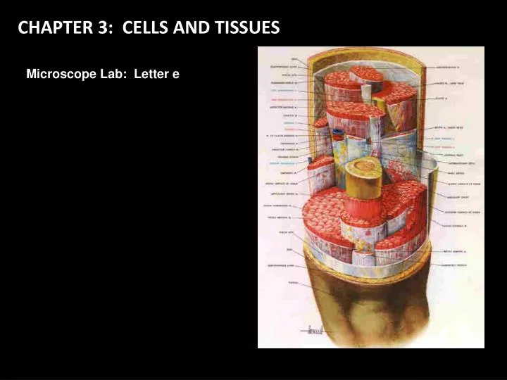
CHAPTER 3: CELLS AND TISSUES. Microscope Lab: Letter e. Anatomy of a Generalized Cell. Did you hear?! QUIZ tomorrow on these structures and functions!. Anatomy of a Generalized Cell: QUIZ. D. A. B. E. F. G. C. H. Q. I. P. O. N. J. M. K. L. VOYAGE INSIDE THE CELL 15 min.

E N D
CHAPTER 3: CELLS AND TISSUES Microscope Lab: Letter e
Anatomy of a Generalized Cell Did you hear?! QUIZtomorrow on these structures and functions!
Anatomy of a Generalized Cell: QUIZ D. A. B. E. F. G. C. H. Q. I. P. O. N. J. M. K. L.
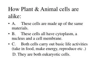
Cell Diversity There are seven primary types of cells found in humans. These types are defined by what they do. REFER to TXT Figure 3.7 pg 65 and descriptions found in text. 1. Cells that connect body parts. A. Fibroblast Elongated shape Fibrous Lots of Rough ER Big Golgi Complex www.footdoc.ca celleng-tech.com www.bioch.ox.ac.uk www.davidlnelson.md
Cell Diversity There are seven primary types of cells found in humans. These types are defined by what they do. REFER to TXT Figure 3.7 pg 65 and descriptions found in text. 1. Cells that connect body parts. B. Erythrocyte Red blood cells Carries oxygen Concave shape provides extra surface area to take on oxygen No organelles www.adamdorman.com
Cell Diversity There are seven primary types of cells found in humans. These types are defined by what they do. REFER to TXT Figure 3.7 pg 65 and descriptions found in text. • Cells that cover and line body organs. • Epithelial Cell • Hexagonal shape • Pack into sheets Intermediate filaments Resists tearing upload.wikimedia.org learn.hamamatsu.com
Cell Diversity There are seven primary types of cells found in humans. These types are defined by what they do. REFER to TXT Figure 3.7 pg 65 and descriptions found in text. 3. Cells that move organs and body parts. A. Skeletal and Smooth muscle cells. Elongated shape Lots of contractile filaments Smooth Muscle Cell class.kmu.edu.tw Skeletal Muscle Cell Flash: Insane Feats of Strength www.blobs.org
Cell Diversity There are seven primary types of cells found in humans. These types are defined by what they do. REFER to TXT Figure 3.7 pg 65 and descriptions found in text. “Empty” Cell 4. Cells that store nutrients. Fat Cells Made of a cell that becomes filled with a lipid droplet. “Filled” Cell Teenage Obesity 30:00 http://player.discoveryeducation.com/index.cfm?guidAssetId=9F3F8962-C7F4-49F5-8D7A-15C990C343D3&blnFromSearch=1&productcode=US# www.blobs.org
Cell Diversity There are seven primary types of cells found in humans. These types are defined by what they do. REFER to TXT Figure 3.7 pg 65 and descriptions found in text. • Cells that fight disease • Macrophage Cells • Contain lots of lysosomes and use pseudopods (false feet) to capture disease units. Flash:Macrophage Cytokine Release relfe.com www.blobs.org
Cell Diversity There are seven primary types of cells found in humans. These types are defined by what they do. • Cells that gather information and control body functions. • Nerve Cells (Neurons) • Have long extensions called Processes that receive and send messages. 2.bp.blogspot.com www.blobs.org images.dpchallenge.com
Cell Diversity There are seven primary types of cells found in humans. These types are defined by what they do. 7. Cells used for reproduction Egg Cells (Oocyte) Female reproductive cell Sperm Cells Male reproductive cell 2.bp.blogspot.com www.blobs.org
Membrane Transport Solution = homogeneous mixture of two or more components. Solute = the substance present in the smallest amount in the solution. Example: Kool-Aid dissolved in Water Water is the solvent. Kool-Aid is the solute. Intracellular Fluid = fluid within the cell Interstitial Fluid = fluid around the outside of the cell Contains nutrients, regulatory substances like hormones, salts, waste products. Each cell pulls what it needs from the interstitial fluid and deposits waste into the interstitial fluid. Intracellular Fluid Interstitial Fluid
Membrane Transport inside cell outside cell • Membrane is made of special kind of lipid • phospholipids • “split personality” • Membrane is a double layer • phospholipidbilayer “attracted to water” phosphate lipid “repelled by water”
Membrane Transport Semi-permeablemembrane • Cell membrane controls what gets in or out • Need to allow some materials — but not all — to pass through the membrane • semi-permeable (semi – partly) • only some materials can get in or out So what needs to get across the membrane? lipids aa O2 H2O salt sugar waste
Membrane Transport inside cell outside cell Crossing the cell membrane • What molecules can get through the cell membrane without doors or help? • fats and oils can pass directly through lipid salt waste but… what about other stuff? sugar aa H2O
Membrane Transport Cell membrane protein channels • Need to make “doors” through membrane • protein channels allow substances in & out • specific channels allow specific material in & out • H2O channel, salt channel, sugar channel, etc. inside cell H2O aa sugar salt outside cell waste
Membrane Transport • Channels are made of proteins • proteins both “like” water & “like” lipids bi-lipid membrane protein channelsin bi-lipid membrane
Membrane Transport Protein channels (cont.) • Proteins act as open doors in the membrane • channels to move specific molecules through cell membrane HIGH Sugar molecules Concentration gradient LOW
Membrane Transport Simple Diffusion • Move from HIGH to LOW Which way will these fat molecules move? fat fat fat inside cell fat fat fat LOW HIGH fat outside cell fat fat fat fat fat fat fat
Membrane Transport FacilitatedDiffusion • Move from HIGH to LOWthrough a channel sugar sugar sugar sugar inside cell sugar sugar LOW Which way will sugar move? HIGH outside cell sugar sugar sugar sugar sugar sugar sugar
Membrane Transport Filtration = movement of water and solutes across a membrane as a result of hydrostatic pressure usually exerted by the blood.
Activetransport • Cells may need to move molecules against concentration gradient • need to pump “uphill” • from LOW to HIGH using energy • Solute PUMP • Requires ATP ATP Notice the direction of Amino Acid movement and the concentrations! Na+ activates the pump. Low High
Membrane Transport Bulk Transport Exocytosis = movement of substances OUT of the cell. Endocytosis = movement of substances INTO the cell.
Osmosis Movement of Water Across Cell Membrane
Membrane Transport • Osmosis • diffusion of water from high concentration of WATER to low concentration of water • across a semi-permeable membrane High Low
Membrane Transport MaintainingHomeostasis • Cell survival depends on balancing water uptake & water loss saltwater balanced freshwater
Cell Processes Mitosis = Division of one cell into two identical cells. Interactive Mitosis http://www.cellsalive.com/mitosis.htm
Cell Processes Protein Synthesis = Processes that use DNA to create proteins.
BODY TISSUES: EPITHELIAL TISSUES Tissues = groups of cells that are similar in structure and function • Characteristics: • 1. Fit closely together. • Held together by desmosomes and tight junctions. • Always have one free edge called the apical surface that is exposed to the body’s exterior or an organ cavity. • Lower surface rests on a basement membrane which it secretes. • Avascular = No blood supply of their own. • Regeneration = ability to make more of themselves. Epithelium:(epithe = covering) tissues of linings, coverings or glands Functions: Protection Absorption Secretion
BODY TISSUES: EPITHELIAL TISSUES Simple Epithelium = one layer of cells Stratified Epithelium = more than one layer of cells Pseudostratified Epithelium = one layer that looks like two. Squamous = flat Cuboidal = short cubes Columnar = tall columns
BODY TISSUES: EPITHELIAL TISSUES Simple Squamous Epithelia • Characteristics: • One layer. • Look like floor tiles. • Found in membranes where filtration or exchange of substances occurs. • Examples: • Lining of air sacs in lungs. • Walls of capilaries. • Serosae = slick membranes lining the body cavity and covering organs. nte-serveur.univ-lyon1.fr Why would this type of tissue need to be thin?
BODY TISSUES: EPITHELIAL TISSUES Simple Cuboidal Epithelia • Characteristics: • One layer. • Look like cubes packed together. • Found in glands and ducts. • Examples: • Salivary glands • Pancreas • Kidney tubules nte-serveur.univ-lyon1.fr
BODY TISSUES: EPITHELIAL TISSUES Simple Columnar Epithelia • Characteristics: • One layer. • Look like columns packed together. • Found in body cavities. • Goblet Cells = produce lubricating mucus. • Examples: • Digestive tract • Mucosae = lining of body cavities that open to exterior.
BODY TISSUES: EPITHELIAL TISSUES Pseudostratified Columnar Epithelia cilia • Characteristics: • One layer. • Looks like two layers because some cells are shorter than others. (pseudo = false) • Functions in absorption and secretion. • Some have cilia. • Examples: • Respiratory tract nte-serveur.univ-lyon1.fr
BODY TISSUES: EPITHELIAL TISSUES Stratified Squamous Epithelia • Characteristics: • Multiple layers. • Most common stratified tissue. • Cells at free edge are squamous. Cells at basement membrane can be columnar or cuboidal. • Found where abuse or friction occurs. • Examples: • Esophagus • Mouth • Outer skin
BODY TISSUES: EPITHELIAL TISSUES Stratified Cuboidal or Columnar Epithelia Characteristics: Multiple layers. Rare. Found in ducts of large glands. Examples: Salivary glands nte-serveur.univ-lyon1.fr
BODY TISSUES: EPITHELIAL TISSUES Transitional Epithelia Characteristics: Multiple layers. Highly modified. Forms lining of a few organs. Examples: Bladder Ureters Urethra nte-serveur.univ-lyon1.fr
BODY TISSUES: CONNECTIVETISSUES Types of Connective Tissue Cartilage tissue: softer than bone, more flexible. Hyaline cartilage = lots of collagen fibers hidden by rubbery matrix that looks like glass (hyalin = glass). Function: Larynx Ribs to breastbone Ends of bones at joints Fetal “bones” qwickstep.com
BODY TISSUES: CONNECTIVETISSUES Types of Connective Tissue Cartilage tissue: softer than bone, more flexible. Elastic cartilage Fibrocartilage = highly compressible cushionlike discs between vertebrae. Function: Vertebral cushioning
BODY TISSUES: CONNECTIVETISSUES Types of Connective Tissue Dense Connective/Fibrous tissue: collagen matrix. Fibroblasts = fiber-forming cells between collagen fibers. Strong, rope-like structures. Tendon = attaches skeletal muscles to bones. Ligament = attaches bones to bones. Function: Connections kentsimmons.uwinnipeg.ca
BODY TISSUES: CONNECTIVETISSUES Types of Connective Tissue Areolar tissue: • Most widely distributed. • Soft, pliable. • Acts as a glue to hold organs together and in their places. • Lamina propria = areolar tissue that underlies all mucosa epithelium. • Looks like mostly space (aerola = small open space) • Function: Cushions and protects • Absorbs waste materials www.tvcc.edu athletictapeinfo.com cheneyhs.org
BODY TISSUES: CONNECTIVETISSUES Types of Connective Tissue Adipose tissue: Commonly called “FAT.” Areolar tissue in which fat cells predominate. Function: Subcutaneous layer under skin. Insulation Protection guaranteedtosleep.com
BODY TISSUES: CONNECTIVETISSUES Types of Connective Tissue Reticular Connective tissue: Associated with reticular cells (similar to fibroblasts). Function: Forms Stroma (framework) that supports free blood cells in lymph nodes, spleen, and bone marrow. www.unomaha.edu www.malecare.com www.helenjaques.co.uk
BODY TISSUES: CONNECTIVETISSUES Types of Connective Tissue Blood: Also called “vascular tissue.” Made of blood cells surrounded by blood plasma (fluid). Function: Transports oxygen, nutrients, water, etc. blog.lib.umn.edu kska.org
BODY TISSUES: MUSCLETISSUES MUSCLE TISSUE • Highly Specialized to contract or shorten. • Elongated. • Also called “muscle fibers.” • Function: Produces movement. www.uic.edu www.deviantart.com www.athleteofficial.com
BODY TISSUES: MUSCLETISSUES Types of Muscle Tissue Skeletal Muscle: • connective sheets • attached to skeleton • voluntarily controlled • cells are long, cylindrical, and multinucleate (many nuclei) • Function: Movement www.uic.edu www.clccharter.org
BODY TISSUES: MUSCLETISSUES Types of Muscle Tissue Cardiac Muscle: • Found only in heart. • Has striations. • Fit together at intercalated disks (like clasped fingers). • Gap junctions allow ions to pass freely from cell to cell which produces electrical beat. • Involuntary muscle. • Function: Pumps blood. cache1.asset-cache.net journals.prous.com ucl.ac.uk academic.brooklyn.cuny.edu
BODY TISSUES: MUSCLETISSUES Types of Muscle Tissue Smooth Muscle: • Also called “visceral muscle.” • No striations. • Found in walls of hollow organs (stomach, blood vessels, uterus, etc.) • Makes cavity of organ smaller or larger. • Peristalsis = wavelike motion that keeps food moving through the digestive system. • Function: Pushes substances through an organ along a specific pathway. Peristalsis http://www.mennellmedia.co.uk/VideoProjects/Peristalsis/Peristalsis.html medsci.indiana.edu