Download
1 / 63
630 likes | 948 Views
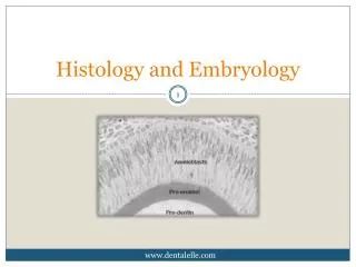
Oral Histology, Embryology and Genetics Spring 2008. Course Syllabus. Oral Mucosa. Mucous Membrane: Moist lining of the gastrointestinal tract, nasal passages and other body cavities that communicate with the exterior

E N D
Oral Histology, Embryology and Genetics Spring 2008
Oral Mucosa Mucous Membrane: Moist lining of the gastrointestinal tract, nasal passages and other body cavities that communicate with the exterior In the oral cavity the lining is called as oral mucous membrane or oral mucosa
Functions of the Oral Mucosa • Protection: Barrier for mechanical trauma and microbiological insults • Sensation: Temperature (heat and cold), touch, pain, taste buds, thirst; • reflexes such as swallowing, etching, gagging and salivating • 3. Secretion: Salivary secretion • 4. Thermal regulation: Important in dogs not in humans
Organization of the Oral Mucosa • 3 types according to FUNCTION: • Masticatory Mucosa: 25% of total mucosa. Gingiva (free, attached • and interdental) and hard palate. Primary mucosa to be in contact with food • during mastication. MASTiCATORY MUCOSA IS USUALLY KERATINIZED. • Lining Mucosa: 60% of total mucosa. Covers the floor of mouth, ventral • (underside) tongue, alveolar mucosa, cheeks, lips and soft palate. • Does not function in mastication and therefore has minimal attrition. • Non-keratinized; soft and pliable. • Specialized Mucosa: 15% of total mucosa. Covers dorsal tongue and • composed of cornified epithelial papillae.
General Features of Oral Mucosa • Separated from the skin by vermillion zone of the lips which is more deeply • colored than rest of the oral mucosa • Factors affecting color of the oral mucosa: • a. Concentration and state of dilation of the blood vessels in • underlying connective tissue • b. Thickness of the epithelium • c. Degree of keratinization • d. Amount of melanin pigmentation • Clinically, color of oral mucosa is very important. For example, inflamed oral • tissues appear red rather than the normal pale pink
How is the oral mucosa different from skin? • Color • Moist surface • Absence of adnexal skin structures such as hair follicles, sweat glands • and sebaceous glands (exception in Fordyce’s disease) • Fordyce’s disease: Sebaceous glands in oral cavity predominantly in upper • lip, buccal mucosa and alveolar mucosa • Presence of minor salivary glands in oral mucosa • Texture of surface: Oral mucosa is smoother than the skin (few exceptions • like dorsal tongue – due to papillae; hard palate – rugae; gingiva – stippling) • Firmness: Oral mucosa varies in its firmness. For example buccal mucosa • and lips are loose and pliable whereas the gingiva and hard palate are firm • so critical clinically while giving injections
Oral Mucosa-Cheek Skin
Fordyce’s Spots • Pale yellow spots • Normal variation • Lips, buccal mucosa, alveolar • mucosa and tonsillar pillar
Structure of Oral Mucosa • Overlying oral epithelium • Underlying connective tissue (lamina propria and submucosa) • In skin called epidermis and dermis Rete ridges/pegs Connective tissue papilla
A: Startum basale B: Startum spinosum C: Startum superficiale A: Epithelium B: Connective tissue C: Salivary gland The oral epithelium is keratinized or non-keratinized stratified squamous epithelium The interface between epithelium and connective tissue is comprised of a structureless layer called basement membrane This interface is irregular and is composed of downward projections of epithelium called rete ridges or rete pegs, and upward projection of connective tissue termed as connective tissue papillae http://dentistry.ouhsc.edu/intranet-WEB/Courses/CELL8002/Home.html
Junction between oral epithelium and lamina propria is more obvious than that between lamina propria and submucosa No muscularis mucosae layer seen in oral mucosa Loose fat and glandular tissue with blood vessels and nerves seen underneath oral mucosa from underneath bone or muscle layer - this layer is termed SUBMUCOSA – provides flexibility In gingiva and hard palate, no submucosa is seen and the lamina propria is directly attached to the periosteum of the underlying bone which provides firm, inelastic attachment – this is called ORAL MUCOPERIOSTEUM
Connective tissue in oral cavity is comprised of salivary glands, sebaceous glands (Fordyce’s disease) and lymphoid tissue (tonsillar tissue) Salivary glandsSebaceous glandsLymphoid tissue (tonsil) High Power view of sebaceous glands www.usc.edu/hsc/dental/opfs/QL/09tn.html http://www.usc.edu/hsc/dental/ohisto/index.html http://dentistry.ouhsc.edu/intranet-WEB/Courses/CELL8002/Home.html
Oral Epithelium Progenitor population: Divide and provide new cells (Proliferation) Maturing population: Undergo differentiation (maturation) Estimated time necessary to replace all the cells in the epithelium: turnover time Skin: 52 to 75 days Gut: 4 to 14 days Gingiva: 41 to 57 days Cheek: 25 days Nonkeratinized epithelium turns over faster than keratininzed epithelium Clinical correlation: Oral ulcers during cancer chemotherapeutic treatment
Types of Oral Epithelium Orthokeratinized stratified squamous epithelium Parakeratinized stratified squamous epithelium Nonkeratinized stratified squamous epithelium
Components of Oral Epithelium Lining Mucosa: Stratum Basale: Basal cell layer comprised of cuboidal cells. Progenitor cells that divide and provide new cells by mitotic division that migrate to the surface to replace cells that are shed. Stratum Spinosum (or intermedium): Cells are oval and represent bulk of the epithelium. Stratum Superficiale: Cells are flat and contain small oval nuclei that are continuously shed.
Histology of Lip • Skin: keratinized stratified squamous • epithelium with adnexal skin structures • Oral Mucosa: Moist-surface, covered by • nonkeratinized stratified squamous epithelium • associated with small round seromucous • glands of the lamina propria. In the • submucosa fibers of orbicularis oris muscle is • noted. • Vermillion zone: Very thin keratinized • epithelium that contains no adnexal skin • structures (can contain sebaceous glands) • What gives the vermillion zone the red color? • Epithelium is thin • Epithelium contains eleidin, which is • transparent • Blood vessels are present near the surface • Eleidin is a semi-fluid clear substance present • in the stratum lucidum of the skin epithelium A: Skin B: Vermillion zone C: Oral (labial) mucosa D: Minor salivary glands http://dentistry.ouhsc.edu/intranet-WEB/Courses/CELL8002/Home.html
Skin of the LipVermillion Zone and Labial Mucosa A: Vermillion zone B: Labial mucosa C: Orbicularis oris muscle A: Sweat glands B: Sebaceous glands C: Hair follicles http://dentistry.ouhsc.edu/intranet-WEB/Courses/CELL8002/Home.html
Soft palate • Nonkeratinized • Highly vascularized so more pink • than hard palate • 2. Lamina propria and submucosa • present (unlike hard palate when • only lamina propria is noted – • mucoperiosteum) • Submucosa contains salivary • glands and muscle soft palate A: Hard Palate B; Soft palate C: Nasal cavity http://dentistry.ouhsc.edu/intranet-WEB/Courses/CELL8002/Home.html
Cheeks (Buccal Mucosa) Similar to lips and soft palate Nonkeratinized stratified squamous epithelium, lamina propria and submucosa Submucosa of cheeks contain fat cells along with lobules of minor salivary glands and muscle fibers
Floor of mouth Ventral surface of tongue Nonkeratinized stratified squamous epithelium, lamina propria and submucosa Epithelium is loosely attached to lamina propria No muscle Nonkeratinized stratified squamous epithelium, lamina propria and submucosa Extremely dense muscle fibers interlacing connective tissue fibers in submucosa
Masticatory Mucosa Epithelium that covers gingiva and hard palate Mucosa is thicker than nonkeratinized because of the keratin layer Stratum basale Startum spinosum Stratum granulosum: Cells contain keratohyaline granules Stratum corneum: Contains thin, flat and nonnucleated cells which are filled with keratin. In contrast to the hard keratin seen in nails and hair, keratin overlying normal masticatory oral mucosa is soft. Keratin is tough, nonliving material that is resistant to friction and impervious to bacterial invasion Same as nonkeratinized epithelium Miller SE. Histology for Pathologists. 3rd edition. LWW. 2007
Types of Keratinized Epithelium Miller SE. Histology for Pathologists. 3rd edition. LWW. 2007 The superficial cells are dead but retain the nucleus in parakeratinized epithelium but the nuclei are lost in orthokeratinized epithelium The rete pegs are long and slender in keratinized epithelium Mucoperiosteum http://dentistry.ouhsc.edu/intranet-WEB/Courses/CELL8002/Home.html
Gingiva and Epithelial Attachment Free or marginal gingiva Attached gingiva attaces with the neck of the tooth by means of junctional epithelium
Histology of Gingiva Thick (250 µm), either orthokeratinized or parakeratinized stratified squamous epithelium with a stippled surface A: Rete pegs B: Conective tissue papilla C: Parakeratin D: Spinous layer In healthy attached gingiva “stippling” is seen which appears as small pits in the epithelium and are due to deep rete pegs. The lamina propria is composed of long narrow papillae which are not highly vascular. No distinct submucosa is noted as the overlying mucosa is directly attached to the underlying periosteum and cementum by collagen fibers http://dentistry.ouhsc.edu/intranet-WEB/Courses/CELL8002/Home.html
Free Gingiva: Keratinized; NOT STIPPLED; bound on inner margin by the gingival sulcus, which separates it from the tooth; bound on its outer margin by the oral cavity; and apically by the free gingival groove. Attached Gingiva: Keratinized; STIPPLED; separated from the alveolar mucosa by the mucogingival junction (groove). Attached to the tooth by junctional epithelium.
In A note the difference in keratinization, thickness of epithelium and ridge pattern. In B stained with an elastic stain, note the presence of elastic fibers in lamina propria in alveolar mucosa. Not much difference in the epithelium is seen between the two types of mucosa.
Dentogingival Junction and Junctional Epithelium Dentogingival junction is the region where the oral mucosa meets the surface of the tooth Very important because it is a weak area in the oral mucosa which is otherwise continuous Bacteria on the surface of the tooth produce toxins that can incite inflammation and damage if it enters into the mucosal tissues Gingival sulcus in healthy individuals is ~ 0.5 to 3 mm (mild inflammation is present- 1.8 mm average) Depth greater that 3mm is considered pathologic; and the sulcus represents periodontal pocket Floor of the sulcus and the epithelium cervical to it is called junctional epithelium which is in contact with the tooth surface (enamel and sometimes cementum) Wall of the gingival sulcus is lined by nonkeratinizing stratified squamous epithelium that is derived from and continuous with the rest of the oral mucosa – oral sulcular epithelium
Junctional Epithelium The epithelium that is attached to the tooth (enamel or sometimes cementum) surface continuous with sulcular epithelium Derived from reduced enamel epithelium of the tooth germ Junctional epithelium consists of flat cells aligned parallel to the tooth surface increasing in thickness from the apex to the crown Attached to enamel by internal basal lamina and to the connective tissue by external basal lamina. Hemidesmosomes are present in both basal laminas.
Epithelial cell turnover in gingiva Similar to all other epithelia, the deeper cells adjacent to the connective tissue undergo cell division to replenish those lost at the surface High rate of cell division Migrate about 2 to 3 cell layers from the tooth surface and then join a main migratory route in a coronal direction, parallel to tooth surface, to be desquamated into the gingival sulcus. Key point: Junctional epithelium readily regenerates from the sulcular epithelium or oral epithelium if it is damaged or surgically excised Connective tissue normally contains plenty of neutrophils which is different than the normal oral mucosa
Col Col (or depression): This is how the gingiva looks in the interdental area. Similar to an outline of a depression or col with buccal and lingual peaks. Col epithelium is identical to junctional epithelium and has the same origin (from dental epithelium) and is also replaced continually by cell division. Not sure if there is any significance that the col is more vulnerable to inflammation, but the incidence of gingivitis is greater interdentally.
A quick note on….. Blood supply to the gingiva: Derived from periosteal vessels in the periosteum of the alveolar process Blood supply to the dentogingival junction: Continuation of interalveolar arteries Nerve supply to the gingiva: terminal branches of periodontal nerve fibers and by branches of the infraorbital and palatine, or lingual, mental, and buccal nerves PLEASE REVIEW TABLE 12-4 AND 12-5 IN THE TEXT BOOK
Histology of Hard Palate Thick orthokeratinized (or parakeratinized in areas) epithelium showing ridges (rugae) Lamina propria shows long papillae with thick dense connective tissue Submucosa is mucoperiosteum with dense collagenous connective tissue attaching directly to periosteum. Contains fat and salivary glands
Specialized Mucosa – Dorsal Tongue Types of papilla: 4 types A: Foliate C: Circumvallate D: Filiform
Filiform papilla: Makes up majority of the papillae and covers the anterior part • of the tongue. They appear as slender, threadlike keratinized projections • (~ 2 to 3 mm) of the surface epithelial cells. These papillae facilitate mastication • (by compressing and breaking food when tongue is apposed to the hard palate) • and movement of the food on the surface of the tongue. The papillae is directed • towards the throat and assist in movement of food towards that direction. • NO TASTE BUDS.
Fungiform papilla: (Fungus-like) These are interspersed between the filiform papilla. • More numerous near the tip of the tongue. Smooth, round structures that appear • red because of their highly vascular connective tissue core, seen through a thin, • nonkeratinized stratified squamous epithelium. Taste buds are usually seen within • the epithelium. Filiform papilla
Foliate Papilla: (Leaf-like). Present on the lateral margins of the posterior tongue. • Consist of 4 to 11 parallel ridges that alternate with deep grooves in the mucosa, • and a few taste buds are present in the epithelium. They contain serous glands • underlying the taste buds which cleanse the grooves.
Circumvallate papilla: (Walled papilla). 10 to 14 in number these are seen along • the V-shaped sulcus between the base and the body of the tongue. Large, ~ 3 mm • in diameter with a deep surrounding groove. Ducts of von Ebner glands (serous • salivary glands) open into the grooves. Taste buds are seen lining the walls of • the papillae.