Download
1 / 16
160 likes | 318 Views
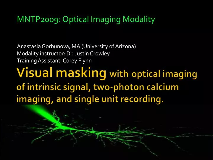
MNTP2009: Optical Imaging Modality. Anastasia Gorbunova, MA (University of Arizona) Modality instructor: Dr. Justin Crowley Training Assistant: Corey Flynn. Visual masking with optical imaging of intrinsic signal, two-photon calcium imaging, and single unit recording. Visual Masking.

E N D
MNTP2009: Optical Imaging Modality Anastasia Gorbunova, MA (University of Arizona) Modality instructor: Dr. Justin Crowley Training Assistant: Corey Flynn Visual masking withoptical imaging of intrinsic signal, two-photon calcium imaging, and single unit recording.
Visual Masking • “Destructive interaction or interference among transient stimuli that are closely coupled in space or time” Forster & Davis (1984) J Exper Psych: Learning, Memory & Language Time DOCTOR 500 ms ####### doctor DOCTOR doctor 50 ms ###### doctor 500 ms R. Fox. (1998). In Handbook of Sensory Physiology. VIII. Perception.
Optical Imaging Modality • Optical imaging of intrinsic signal - wide spatial field - temporal resolution slow hemodynamic dependent faster light scattering properties of tissue • Single unit recordings - best temporal resolution • Two-photon Calcium imaging - distinct neurons, fine time course
Stimuli 1. 2. Time Time 4. 3. Time Time
Stimuli 5. 6. 8. 7. Time Time
Intrinsic Signal OI • Exposed visual cortex of a ferret (P80-130) • Illumination by ~700 nm light • Visual stimulation (4 degree bars) • CCD camera (100 ms data frames) • Computer digitization 8000 ms Courtesy of David E. Whitney
Experiment 2: F09-051 1 Block ISB 3 s Time Blank stimulus 8.1 s ISB 3 s 8 s Stimulus 2 0.1 s Inter-Stimulus Blank 3 s 8 s Stimulus 1 0.1 s - =
Experiment 2: F09-051 8 s 8 s 8 s 100 ms 100 ms 100 ms
Experiment 2: F09-051 8 s 8 s 8 s 1 frame: 500 ms 100 ms 100 ms 100 ms
Extracellular Single Unit Recordings • Exposed visual cortex of a ferret (P80-130) • Carbon fiber electrode • 20kHz signal digitization
Extracellular Single Unit Recordings V B V B H H SQP SNP SNP SQP H-V H-B H-V V-H H-B V-H V-B V-B H, horizontal grating; B, blank; V, vertical grating; SQP, square-wave plaid; SNP, sine-wave plaid
2-photon calcium imaging • Oregon green BAPTA-1AM (labels neurons and astrocytes) • Sulphorhodamine-101 (labels astrocytes) • Microscope: Zeiss LSM 510 Meta NLO with MIRA900 Ultrafast Ti:Sapphire laser • 20x NA1 objective
2-photon calcium imaging 3 cells: ΔF/F for 700 frames normalized over 10 blocks
Conclusions • It is possible to study masking in animal models using simple visual stimuli • Very brief presentation of static gratings produces sufficient activation detectable by optical imaging of intrinsic signal • Orthogonal mask disrupts that activation
Acknowledgements Dr. Justin Crowley Corey Flynn David Whitney Sruthi Chintakunta Alberto Vazquez Dr. Seong-Gi Kim Dr. Bill Eddy Tomika Cohen Rebecca Clark Thank you!