Download
1 / 64
660 likes | 870 Views
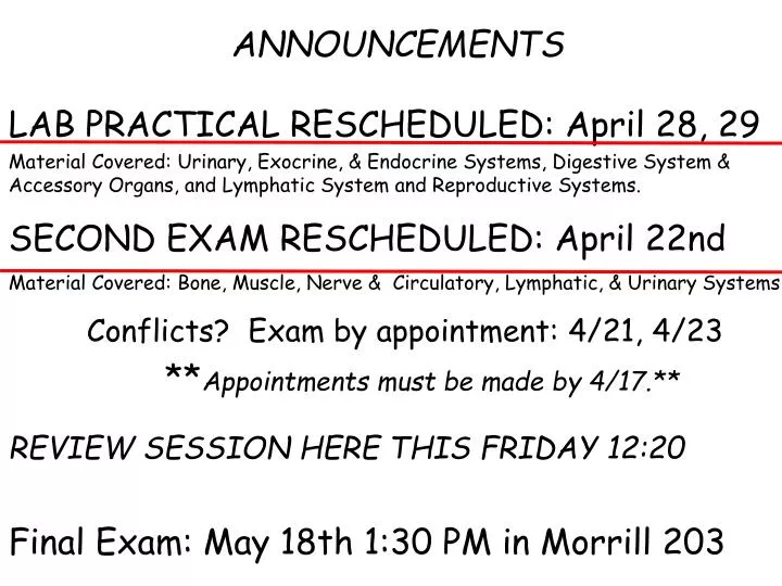
ANNOUNCEMENTS. LAB PRACTICAL RESCHEDULED: April 28, 29 Material Covered: Urinary, Exocrine, & Endocrine Systems, Digestive System & Accessory Organs, and Lymphatic System and Reproductive Systems. SECOND EXAM RESCHEDULED: April 22nd

E N D
ANNOUNCEMENTS LAB PRACTICAL RESCHEDULED: April 28, 29 Material Covered: Urinary, Exocrine, & Endocrine Systems, Digestive System & Accessory Organs, and Lymphatic System and Reproductive Systems. SECOND EXAM RESCHEDULED: April 22nd Material Covered: Bone, Muscle, Nerve & Circulatory, Lymphatic, & Urinary Systems Conflicts? Exam by appointment: 4/21, 4/23 **Appointments must be made by 4/17.** REVIEW SESSION HERE THIS FRIDAY 12:20 Final Exam: May 18th 1:30 PM in Morrill 203
The Lab Project • GOAL: To work as a team to analyze an organ with embedding & sectioning & immunohistochemistry. • To prepare: • Identify your group members. • Identify 3 organs you would like to work on. Possible organs are: liver, brain, bladder, lung, pancreas (?), skeletal muscle, stomach, intestine, kidney, uterus. • Choose 2 antibodies to stain frozen sections. All sections will be stained with DAPI, a marker of cell nuclei. Choose 2 other markers from the list under Review Materials. Label one with a red fluorochrome and one with a green fluorochrome.
LAB SCHEDULE Week of April 13: Learn to frozen section organs. Week of April 20: Complete frozen sections. Week of April 27: Lab Practical and learn to section embedded material. Week of May 4: Immunohistochemistry. Complete sectioning & staining of embedded material. In class, May 11: Share results, discuss interpretation. Each student writes own report according to the guidelines provided on the Review Materials page. May 18: Lab Project report due at Final Exam.
*BIOLOGY SENIORS* Join us for lunch! 12 Noon Wednesday, May 13th Lawn, Durfee Conservatory
Digestive System Alimentary Canal and Associated Organs Mouth Tongue Esophagus Teeth Stomach Salivary Glands Small Intestine Pancreas Large Intestine Liver Gall Bladder
Alimentary Canal Barrier: between internal and external environments Motility: movement of food Secretion: enzymes, mucous, acid, antibodies Absorption: products of digestion Immunological Defense: site of lymphatic tissue
Alimentary Canal General Structure from Esophagus ---> Anus Mucosa: Epithelium Lamina Propria Muscularis Mucosa (smooth muscle) Submucosa: Dense irregular connective tissue Muscularis externa: Two layers of smooth muscle Serosa:simple squamous epithelium, connective tissue
Barrier- Epithelium Oral Cavity: parakeratinized epithelium- most superficial cells do not lose nuclei tongue, gums, hard palate Connective tissue papilla
Barrier- Epithelium Esophagus: stratified squamous epithelium Small and Large Intestine- tight junctions between columnar cells of simple epithelium
Immunological Defense Tonsils: ring of lymphatic tissue (lymphatic nodules or follicles) at entrance to respiratory and digestive tracts micro.magnet.fsu.edu/optics/intelplay/gallery...
Adenoids: lymphatic tissue located high on the posterior wall of the pharynx. • - similar to tonsils • clear antigens from air • - reduced in adults • - can be enlarged / inflamed • SYMPTOMS: • mouth breathing • snoring • bad breath • chronic runny nose • sleep apnea • pulmonary hypertension • right-sided heart failure
Immunological Defense Gut-associated lymphatic tissue (GALT): diffuse lymphatic tissue and lymphatic nodules in lamina propria of small and large intestine Striking in Appendix and Ileum=> Peyer’s Patches MALT=Mucous associated lymphatic Tissue
Immunological Defense Plasma Cells secrete a special form of antibody, ==> secreted IgA -Dimeric -Linked via J chain and secretory component -More stable -More resistant to enzymatic digestion -in saliva, milk, and mucous membranes of respiratory and digestive tracts
Possible modes of defense mediated by IgA binding to its receptor, pIgR, (the secretory component , SC). pIgR-driven export of dimeric IgA with J chain (IgA+J) Neutralization of infecting virus and transport of viral products to the lumen. Intracellular neutralization of endotoxin (LPS) from Gram-negative bacteria. Clearance of antigen (Ag) that has breached the mucosal barrier. From Trends Immunol. 2004, 25:150-57.
Immunological Defense Peyer’s Patches Lymph nodules capped by specialized epithelial cells, =>M Cells www.bu.edu/histology/p/12001oba.htm
M Cells • - Follicle-Associated Epithelium (FAE): epithelial cells associated with lymph nodules of MALT • look for absence of goblet cells over Peyer’s Patch • apical surface microfolds rather than microvilli • - connected to neighbors with tight junctions
M Cells • have extensive inpocketings of basal membrane containing T and B lymphocytes www.rcai.riken.go.jp/eng/group/epi/
M Cells: specialized for transepithelial transport: deliver intact foreign antigens and microorganisms from lumen to immune cells
Motility Muscularis Mucosa: 2 layers of smooth muscle inner-circular, outer-longitudinal responsible for moving the mucosa
Motility Muscularis Externa: mixes, propels contents of lumen 2 thick layers of smooth muscle inner layer=> circularly-oriented layer -tight spiral outer layer=>longitudinally-oriented layer -loose spiral Between muscle layers- Nervous innervation Myenteric plexis (Auerbach’s plexis)
Motility MUSCULARIS EXTERNA EXCEPTIONS: Striated muscle in proximal esophagus (upper 1/3) and anus
MUSCULARIS EXTERNA EXCEPTIONS: Teniae Coli: 3 thickened bands of longitudinal layer large intestine-
Secretion • carried out by epithelial cells and associated glands • secretions include: • Antibodies: IgA • Lubrication substances • Aid for digestion: hydrochloric acid & enzymes • Hormones • Water • secretions from salivary glands, stomach, small and large intestine
Anatomy of the Stomach 3 regions: Cardiac Pyloric Fundic Rugae: longitudinal folds or ridges on inner surface
Anatomy of the Stomach 3 regions: Cardiac Pyloric Fundic Rugae: longitudinal folds or ridges on inner surface Simple columnar epithelium
Each stomach region • has distinctive glands. • Cardiac glands • Pyloric glands • Fundic glands • -gastric pits • -isthmus • cell replication • -neck • -base or fundus
Anatomy of the Small Intestine 3 components: Duodenum, Jeunum, Ileum - Plicae circularis - Villi - Microvilli - Simple columnar epithelium
Secretion / Digestion / Absorption epithelial cells and associated glands salivary glands pancreas gall bladder stomach small & large intestine Secretions include: antibodies: IgA lubricants hydrochloric acid digestive enzymes hormones water
Secretion / Digestion / Absorption - initiated in mouth - stomach lumen - completed in small intestinal lumen - aided by HCl from stomach - amylase from saliva & stomach - pancreatic enzymes - enzymes in glycocalyx of small intestine - aided by bile from gall bladder
Secretion / Digestion / Absorption across epithelium of small intestine and large intestine 10.
Lubrication: Mucous Secretions Esophagus- Lubrication and protection from regurgitation of acidic stomach contents Stomach- surface mucous cells; mucous protects from abrasion, contains bicarbonate; protects mucosa from acidic stomach contents (chyme) Small Intestine- goblet cells, # increases from duodenum=> ileum Large Intestine- goblet cells, # increases toward rectum
Mouth Stomach Small Large IntestineIntestine Carbohydrate Protein Lipids Nuclei Acids Digestive Secretions Digestive Secretions from Pancreas Absorption Bile from gall bladder
Specialized Cells for Stomach Secretion Surface Mucous Cells: gastic pit and neck of gastric gland PAS stain for carbohydrates millette.med.sc.edu/Lab%201%20pages/introduct...
Specialized Cells for Stomach Secretion Parietal (Oxyntic) Cells: - neck & deep parts of fundic glands - release HCl and intrinsic factor (B12 absorption) - large - triangular - acidophilic
Parietal (Oxyntic) Cells Anti-parietal cell antibody
Parietal (Oxyntic) Cells HCl Synthesis: H+ and Cl- ions pumped into intracellular canalicular system, HCl formed http://www.mfi.ku.dk/ppaulev/chapter22/images/22-10.jpg
Specialized Cells for Stomach Secretion Chief Cells: deep in fundic glands, protein-secreting, lots of RER, basophilic, zymogen granules Secrete pepsinogen HCl Pepsinogen---------> Pepsin
Specialized Cells for STOMACH Secretion Enteroendocrine cells: small - more common in gland base - pale, vesicles don’t fix well - may not reach lumen, but sample lumenal contents with microvilli -release variety of hormones into blood
Small Intestine Increased Surface Area: Plicae circularis, Villus, Intestinal Gland (Crypts of Lieberkuhn) Villus: Capillary Lacteal (lymphatic capillary) Smooth muscle
Specialized Cells of the Small Intestine Enterocytes (intestinal absorptive cells) Paneth cells- secrete antimicrobial substances Enteroendocrine cells- release hormones M cells- dome cells cap lymphatic nodules Goblet cells- mucous secreting
Enterocytes (intestinal absorptive cells) Tall columnar cells Microvilli=>striated border Epithelial specializations -Terminal web - Tight junctions Secrete Digestive Enzymes
Paneth Cells • - base of intestinal glands • large • intense acidophilic granules • phagocytose bacteria • secrete lysozyme- digests • bacterial cell wall
Large Intestine Simple columnar epithelium Absorption of water and electrolytes Columnar absorptive cells Crypts of Lieberkuhn Goblet cells www.kumc.edu/.../histoweb/gitract/gi21.htm