Download
1 / 40
850 likes | 2.35k Views
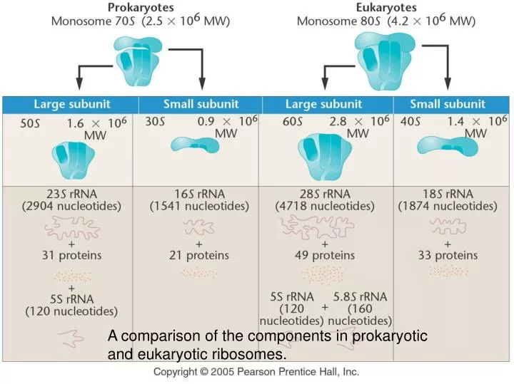
A comparison of the components in prokaryotic and eukaryotic ribosomes. . Cloverleaf Model of Transfer RNA. A three-dimensional model of transfer RNA. TRANSLATION: making proteins

E N D
A comparison of the components in prokaryotic and eukaryotic ribosomes.
TRANSLATION: making proteins http://www.brookscole.com/chemistry_d/templates/student_resources/shared_resources/animations/protein_synthesis/protein_synthesis.html
Elongation of the growing polypeptide chain during translation
Polyribosomes as seen under the electron microscope. They were taken from giant salivary gland cells of the midgefly, Chironomus thummi. Note that the nascent polypeptide chain is apparent as it emerges from each ribosome. Its length increases as translation proceeds from left (5’) to right (3’) along the mRNA.
Each amino acid has two abbreviations; that is, alanine is designated either ala or A (a universal nomenclature).
secondary level (a) The right-handed alpha helix, which represents one form of secondary structure of a polypeptide chain. (b) The beta-pleated sheet, an alternative form of secondary structure of polypeptide chains.
tertiary level The tertiary level of protein structure in a respiratory pigment, myoglobin. The bound oxygen atom is shown in red.
quaternary level hemoglobin The quaternary level of protein structure as seen in hemoglobin. Four chains (two alpha and two beta) interact with four heme groups to form the functional molecule.
hemoglobin hemoglobin
sickle-cell anemia normal hemoglobin two sickle-cell hemoglobin molecules stuck together
sickle-cell anemia Investigation of hemoglobin derived from HbAHbA and HbSHbS individuals by using electrophoresis, protein fingerprinting, and amino acid analysis. Hemoglobin from individuals with sickle-cell anemia (HbSHbS) (a) migrates differently in an electrophoretic field, (b) shows an altered peptide in fingerprint analysis, and (c) shows an altered amino acid, valine, at the sixth position in the Beta chain. During electrophoresis, heterozygotes (HbAHbS) reveal both forms of hemoglobin.
Metabolic pathway involving phenylalanine and tyrosine. Various metabolic blocks resulting from mutations lead to the disorders phenylketonuria, alkaptonuria, albinism, and tyrosinemia.
Hydroxylation of phenylalanine to tyrosine by the enzyme phenylalanine hydroxylase.
phenylalanine hydroxylase
Induction, isolation, and characterization of a nutritional auxotrophic mutation in Neurospora. (a) Most conidia are not affected, but one conidium (shown in red) contains the mutation. In (b) and (c), the precise nature of the mutation is established and found to involve the biosynthesis of tyrosine.