Download
1 / 25
270 likes | 1.35k Views
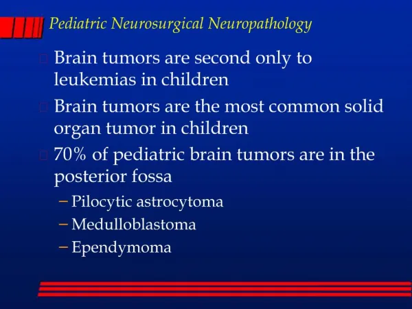
Pediatric Neurosurgical Neuropathology. Brain tumors are second only to leukemias in children Brain tumors are the most common solid organ tumor in children 70% of pediatric brain tumors are in the posterior fossa Pilocytic astrocytoma Medulloblastoma Ependymoma.

E N D
Pediatric Neurosurgical Neuropathology • Brain tumors are second only to leukemias in children • Brain tumors are the most common solid organ tumor in children • 70% of pediatric brain tumors are in the posterior fossa • Pilocytic astrocytoma • Medulloblastoma • Ependymoma
CNS tumors: pediatric vs. adult • Adults: 70% of tumors are supratentorial • meningioma • pituitary adenoma • High grade astrocytoma • Anaplastic astrocytoma (grade III) • Glioblastoma multiforme (grade IV astrocytoma) • Pediatric: 70% in posterior fossa • pilocytic astrocytoma (cerebellar astrocytoma) • medulloblastoma
Brain tumors: intro • Intracranial neoplasms • Primary • Secondary • Metastatic • Local invasion • Tumors of the spinal cord
Primary brain tumors: intro • Primary brain tumors are rare • 2.5% of all cancer deaths • Second most common type of tumor in children • There are over 100 different brain tumors • Most common types • Astrocytomas (adults and children) • Grades I-IV • Medulloblastomas (in children) • also known as: primitive neuroectodermal tumor-PNET • Meningiomas (adults) • Pituitary adenomas (adults)
Clinical presentation • Clinical symptoms depend upon: • Age, location, and type of tumor and grade • Symptoms may include: • Increased intracranial pressure • secondary to obstruction of CSF at aqueduct • hydrocephalus (infants), headache, papilledema, vomiting • seizures • focal neurological deficits • hormonal changes (pituitary adenoma) • visual changes (diplopia, field defects) • Pituitary adenoma - pressure on optic chiasm
CNS tumors: diagnosis • Symptoms prompt neuroimaging • CT and MRI • intra-axial vs. extra-axial • Location of tumor • contrast enhancement • typical of high grade • also in some low grade, i.e., pilocytic astrocytomas
CNS tumors: location • Extra-axial • meningiomas • Cerebral hemispheres • grade II-III astrocytomas, GBM • Crossing corpus callosum - GBM • optic nerve - pilocytic astrocytoma • (in Neurofibromatosis Type 1) • Sella - Pituitary adenoma • Peri-III ventricle - Pilocytic astrocytoma, GBM
CNS tumors: location • posterior fossa (in children) • pilocytic astrocytoma • medulloblastoma • brainstem (pons) • pontine glioma (astrocytoma) • spinal cord • low-grade astrocytomas (grade I and II)
Pilocytic astrocytomas • Most common in children • Grade I astrocytoma • Cerebellum (posterior fossa), optic nerve • Thalamic, spinal cord, cerebral • Discrete, well circumscribed mass • Often with associated cystic area • Contrast enhancing • Histologic appearance: • Biphasic: piloid cells and microcystic areas • Rosenthal fibers • no mitoses
Pilocytic astrocytomas Rosenthal fibers Tumor of cerebellum, often with cyst, biphasic, Rosenthal fibers, piloid cells (enlongated bipolar) biphasic Piloid cells cyst
Astrocytoma - high grade • Astrocytoma grade II and III are very, very rare in the pediatric population • Grade IV - glioblastoma multiforme • Most common astrocytoma in adults • Only about 10% of astrocytomas in children • Diffusely infiltrating tumor of cerebral hemispheres • Contrast enhancing tumor • Histological appearance: • Densely cellular, with marked nuclear pleomorphism • Numerous mitoses • Endothelial proliferation • Necrosis with pseudopallisading
Glioblastoma (grade IV) • Less common in children than adults, typical pathology (necrosis with pseudopallisading)
Pontine glioma Diffuse expansion of pons, usually high grade astrocytoma (III-IV)
Medulloblastomas • PNET of posterior fossa in children • Histologic appearance: • Densely cellular “small blue cell tumor” • Numerous mitoses • Apoptotic (karyorrhectic) cells • Endothelial proliferation • Necrosis • neuronal or glial differentiation • Homer Wright rosettes • GFAP positive cells
Medulloblastoma • Mass arising in roof of fourth ventricle • Homer Wright rosettes
Ependymoma • Mass arising in floor of fourth ventricle • Perivascular pseudorosettes
Meningiomas • Discrete non-invasive tumor • Extra-axial, pushes into brain • Attached to dura • Hyperostosis or invasion of skull common • Histologic appearance: • Fibroblastic or menigothelial cells • Meningothelial whorls • Psammoma bodies • Rare in children, may be intraventricular (lateral ventricles)
Meningiomas Extra-axial tumor, meningothelial cells, whorls and psammoma bodies
Ganglioglioma Cerebrum, cervicomedullary, often with cystic component Increased numbers of neurons (some binucleate) and increased glial cells (usually astrocytic)
Craniopharyngioma • Heterogeneous, cystic mass in suprasellar region • Basiloid layer, stellate reticulum, “wet” keratin, often calcified
Choroid plexus papilloma • Lateral ventricle in children (fourth ventricle in adults)
Germ cell tumors Germinoma Teratoma • Pineal - 99% males, most are germinomas • Suprasellar - often mixed germ cell tumor, 50% female • Teratomas are rare
Metastatic tumors • The most common “brain” tumor in adults is metastatic • Metastatic tumors are rare in children • The most common metastatic tumor in children is osteosarcoma • Local extension of malignant tumors of vertebral bodies (Ewing’s sarcoma) or paravertebral soft tissues (neuroblastoma) are not uncommon
Other tumors • Subependymal giant cell astrocytoma (SEGA) • Intraventricular tumor in Tuberous sclerosis • Desmoplastic infantile ganglioglioma (DIG) • Superficial cerebral tumor in infants • Dysembryoplastic neuroepithelial tumor (DNET) • Hamartomatous lesion associated with seizures • Atypical teratoid rhabdoid tumor (ATR, AT/RT) • Infants, posterior fossa, very malignant • Eosinophilic granuloma • A type of Langerhans cell histiocytosis • Single discrete osteolytic lesion in skull • Meningioangiomatosis • Hamartomatous superficial cerebral lesion associated with seizures
Hereditary syndromes • Neurofibromatosis type I • Café-au-lait spots • Dermatofibromas, multiple • optic nerve astrocytomas, bilateral • plexiform neurofibroma • Malignant peripheral nerve sheath tumor • Neurofibromatosis type II • bilateral acoustic neuroma • multiple meningiomas • ependymomas