Download
1 / 19
340 likes | 957 Views
Magnetic Resonance Imaging (MRI). Steven McLellan. What is MRI?. Produces very clear, detailed pictures of the organs and structures in the body It is a form of medical imaging that uses no Ionizing radiation

E N D
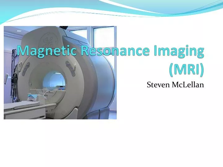
Magnetic Resonance Imaging(MRI) Steven McLellan
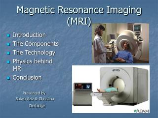
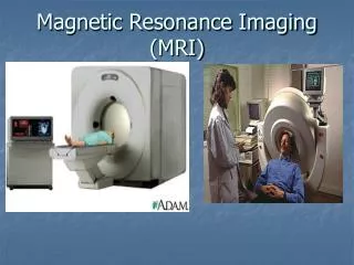
What is MRI? • Produces very clear, detailed pictures of the organs and structures in the body • It is a form of medical imaging that uses no Ionizing radiation • MRI makes use of the property of Nuclear magnetic resonance (NMR) to image nuclei of atoms inside the body.
History • The first MR image was published in 1973 • The first studies performed on humans were published in 1977 • Created by Dr. Raymond V. Damadian, Dr. Larry Minkoff and Dr. Michael Goldsmith • In 2003, The 2003 Nobel Prize in Physiology or Medicine was awarded to Paul C Lauterbur and Peter Mansfield • Made new MR imaging techniques • Faster and more efficient
Common Uses • Physicians use the MR examination to help diagnose or monitor treatment for conditions such as: • Tumors and other cancer related abnormalities. • Certain types of heart problems. • Blockages or enlargements of blood vessels • Diseases of the liver, such as cirrhosis, and that of other abdominal organs. • Diseases of the small intestine, colon, and rectum
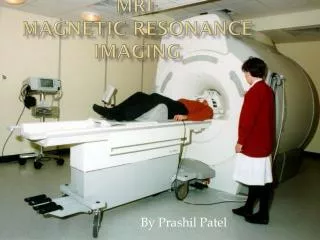
How does it work? • An MRI machine uses a powerful magnetic field to align the magnetization of some atoms in the body. • radio frequency fields systematically alter the alignment of this magnetization • This causes the nuclei to produce a rotating magnetic field detectable by the scanner • This information is recorded to construct an image of the body.
How does it work? • Images are constructed when protons in different tissues return to equilibrium state at different rates. • Five variables effect these rates • Spin Density: Concentration of nuclei in tissue processing in a given region under a magnetic field. • T1: Longitudinal relaxation time • T2: Transverse relaxation time • Flow: Shows blood flow, CSF flow • Spectral Shifts: Angle/zoom the picture is taken from.
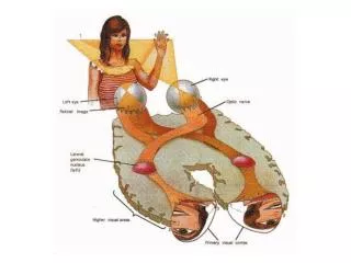
Basic MRI Scans • T1-weighted: Differentiate fat from water • Water is Darker, fat is brighter • Provide good gray matter/white matter contrast in brain. • T2-weighted: Differentiate fat from water • Fat shows darker, and water lighter. • Good for imaging edema • Abnormal accumulation of fluid beneath the skin or in one or more cavities of the body
Specialized MRI Scans Diffusion MRI • Measures diffusion of water through biological tissues. • Diffusion may be anisotropic (unequal physical properties along different axes) • Diffusion Tensor Imaging (DTI) • Examine areas of neural degeneration and demyelination in diseases such as Multiple Sclerosis (MS)
Specialized MRI Scans Magnetic resonance angiography (MRA) • Generates pictures of arteries. • Evaluates the arteries of the neck and brain, the thoracic and abdominal aorta, the renal arteries, and the legs • Uses gadolinium injection as paramagnetic contrast agent • Magnetic resonance venography (MRV) is a similar procedure that is used to image veins.
Safety Risks • MRI’s create up to 120dB • Equivalent to jet engine at take off. • Contraindications: • Pacemakers, Vagus Nerve Stimulators, implantable defibrillators, insulin pumps, deep brain stimulators • Any electronic or magnetized foreign bodies (surgical prosthesis) • Peripheral nerve stimulation (PNS) • Rapid switching on and off of the magnetic field gradients is capable of causing nerve stimulation
During Procedure • People hold the part of their body being scanned motionless for 30-60 minutes. • Procedure is done in multiple parts. • Takes time to switch between different scans and fields of view.
Future? • More detailed images • All MRIs use color? • Better pictures of bone structures • Shift from x-rays and CT scans to MRI • New Scanning sequences
Citations • Dyson, Sue J. Magnetic Resonance Imaging. Philadelphia, PA: Elsevier, 2007. Print. • Hashemi, Ray H., William G. Bradley, and Christopher J. Lisanti. MRI: the Basics. Philadelphia, PA: Lippincott Williams & Wilkins, 2010. Print. • "Magnetic Resonance Imaging (MRI) - Body." RadiologyInfo - The Radiology I nformation Resource for Patients. Radiological Society of North America, Inc., 15 Mar. 2010. Web. 06 Mar. 2011. <http://www.radiologyinfo.org/en/info.cfm?pg=bodymr>. • Radiology". http://radiology.rsna.org/content/204/1/272.long. Retrieved 2 August 2010. • Westbrook, Catherine. MRI. John Wiley & Sons, Incorporated, 2009. Print.