Download
1 / 13

E N D
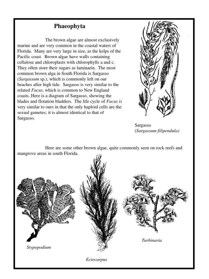
Phaeophyta The brown algae are almost exclusively marine and are very common in the coastal waters of Florida. Many are very large in size, as the kelps of the Pacific coast. Brown algae have walls containing cellulose and chloroplasts with chlorophylls a and c. They often store their sugars as laminarin. The most common brown alga in South Florida is Sargasso (Sargassum sp.), which is commonly left on our beaches after high tide. Sargasso is very similar to the related Fucus, which is common to New England coasts. Here is a diagram of Sargasso, showing the blades and flotation bladders. The life cycle of Fucus is very similar to ours in that the only haploid cells are the sexual gametes; it is almost identical to that of Sargasso. Sargasso (Sargassum filipendula) Here are some other brown algae, quite commonly seen on rock reefs and mangrove areas in south Florida. Turbinaria Stypopodium Ectocarpus
Rhodophyta The red algae are characterized by chlorophyll a and red pigments, called phycobilins, in their chloroplasts. These multicellular algae appear reddish in appearance and are extremely common in marine waters in south Florida. Their walls contain cellulose and quite often accumulate calcium carbonate. They store sugars as Floridean starch. Red algae take on a variety of forms, often as flat blades or as highly branched “trees”. Here are some examples of red algae often seen in south Florida. Grateloupia Dasya Spyridia Cryptarachne Porphyra
Chlorophyta The green algae are the ancestors of terrestrial plants. They have chloroplasts with chlorophylls a and b, the same as in land plants. They also have walls of cellulose, and they store sugars as starch. They vary dramatically in size, from single and motile cells, to filaments, to much larger blades and branched structures. Some accumulate calcium carbonate in their walls and are quite tough. Others, as Ulva – the sea lettuce, are very fragile. Single celled and filamentous algae will be common in ponds and periphyton. The larger marine algae will be encountered in coastal waters throughout south Florida. For examples of the single-celled and filamentous algae look at the illustrations in the description of periphyton. Here we give some examples of algae commonly seen in coastal marine waters. Halimeda Udotea Ulva Codium Caulerpa
Phyrophyta These single celled algae are commonly known as the dinoflagellates. They are important in food webs in tropical marine waters. These algae are distinguished by plates in their cell walls, and they generally are motile. The toxic red tides that occur on the Gulf Coast are blooms of dinoflagellates. Ciguatera, a toxin in some reef fish, is due to the passage of a product of a dinoflagellate in the food web. Some dinoflagellates are illustrated below.
Kingdom Protistsa – the protozoans Protozoans (proto = first, zoan = animal) are among the most versatile of all organisms on earth. Protozoa, however, like algae, is a descriptive term rather than a taxonomic group. Protozoans have an animal-like lifestyle, which means they are active consumers and not photosynthetic. Typically, protozoan have food vacuoles to enclose food particles for digestion and contractile vacuoles to expel excess water. Their single cells employ a variety of features for motility and occupy virtually every microhabitat. The Amoebas Amoebas occur throughout the world in marine, freshwater, and terrestrial Environments. The unifying characteristic of this phylum is the presence of pseudopods, which are moveable extensions of cytoplasm used for locomotion and gathering food. Amoebas lack flagella, and most reproduce asexually
Amoeba is a genus among many organisms commonly called amoebas, and has a structure and physiology typical of most amoeboid genera (refer to picture on previous page). Amoebas are phygocytic, meaning they engulf food particales and form a food vacuole surrounded by a membrane. They then secrete enzymes into the food vacuole for intracellular digestion. A contractile vacuole maintains the cell’s water balance by accumulating and expelling excess water. Amoebas Procedure - observe Amoeba movement and structure • Use a dissecting microscope to examine a culture of living Amoeba. Locate individuals on the bottom. • Prepare a wet mound to living Amoebas by using an eyedropper to remove a few drops from the bottom of the culture of organisms. • Put the drops in a depression slide if one is available or use a standard slide. • Cover the preparation with a coverslip and examine under low power (10x). Soon the Amoeba should move by extending their pseudopods. • If nutrient broth is available, add a drop to the preparation and observe the Amoeba’s response. Questions: • Can you detect moving cytoplasm in the extending pseudopods of the Amoeba? • What do you suppose the living Amoeba is moving towards or away from? • How does the Amoeba respond t nutrient broth? • About how long would it take an Amoeba to move across the field of view on low power? Calculate the rate of movement in mm/hr • Why is a contractile vacuole of a protozoan often more difficult to see than a food vacuole? • Why would excess water tend to accumulate in Amoeba?
Phylum Ciliophora (ciliates) More than 8000 species of ciliate have been described, all having characteristically large numbers of cilia. Most ciliates also have two types of nuclei: micronuclei and macronuclei. A micronucleus divides by mitosis and contains the genetic information of the cells in normally shaped chromosomes. As many as 80 micronuclei may occur in a single cell. The single macronucleus in a cell contained multiple copies of DNA divided into small pieces. The macronucleus replicates by elongating and constricting. Macronuclei are essential for routine cellular functions. Paramecium This free-living fresh water genus is widely studied and easily observed. Paramecium, like most ciliates, undergoes a sexual process called conjugation. During conjugation, individuals from two different strains align longitudinally and exchange nuclear material. This exchange seems to rejuvenate the individuals and is usually followed by frequent mitosis. Asexual reproduction is more common than conjugation and includes mitosis of the micronucleus and transverse fission of the macronucleus and cell body. Procedure - Observe living Paramecium • Prepare a wet mount from a culture of living organisms. • Add a drop of methylcellulose to your wet mount to slow down the Paramecium and make it easier to examine. Questions: • Are the cilia visible on living Paramecium? • Does Paramecium rotate as it moves? • How does the movement of Paramecium compare with that of Amoeba? Figure of a paramecium
Fungi Fungi are among the most common and important groups of organisms. They are basically filamentous strands of cells that secrete enzymes and feed on the organic material on which they are growing. That organic matter might be humus in the soil where mushrooms grow or on stale bread. It may grow between your toes inhabited by athlete’s foot fungus or on a decaying animal on the forest floor being decomposed by fungi digesting the animal’s dead tissue. Fungi not only cause disease: they are important decomposers that recycle nutrients from dead organisms. Fungi which feed on dead organic matter are called saprophytes. The fundamental organization of all fungi is a tube consisting of a series of cells with one or two nuclei (or sometimes with no cell walls partitioning the tube), which is called a hypha (or plural, hyphae). They typically grow together, sometimes into a large organ like a mushroom, as a mycelium. A mycelium can permeate soil, water, or living tissue. In all cases, the hypae of a fungus secrete enzymes for extracellular digestion of the organic substrate. Then the mycelium and its hypae absorb the digested nutrients. For this reason, fungi are called absorptive heterotrophs. Hypae of some species of fungi have crosswalls called septa that separate cytoplasm and nuclei into cells. Hypae of other species have incomplete or no septa (i.e. are aseptate) and therefore are coenocytic (multinucleate). Notably, the cell walls of fungi are usually not cellulose but are made of chitin, the same polysaccharides that comprises the exoskeleton of insects and crustaceans.
Asexual Reproduction Fungi commonly reproduce asexually by mitotic production of haploid vegetative cells called spores in sporangia, conidophores, and other related structures. Spores are microscopic and surrounded by a covering well suited for the rigors of distribution into the environment. Budding and fragmentation are two other methods of asexual reproduction. Budding is mitosis with an uneven distribution of cytoplasm, and is common in yeasts. After budding, the cells with the lesser amount of cytoplasm eventually detach and matures into a new organism. Fragmentation is the breaking of an organism into one or more pieces, each of which can develop into a new individual. Sexual Reproduction The sexual life history of fungi includes the familiar events of vegetative growth, genetic recombination, meiosis, and fertilization. However, the timing of these events in unique to fungi. Fungi reproduce sexually when hypae of two genetically different individuals of the same species encounter each other. Four key features of the fungi life cycle are: Nuclei of a fungal mycelium are haploid during most of the life cycle Gametes are produced by mitosis and differentiation of haploid cells rather than directly from meiosis of diploid cells Meiosis quickly follows formation of the zygote, the only diploid stage Haploid cells produced by meiosis are not gametes; rather, they are spores that grow into a mature haploid organism. Reproductionin Fungi
The Zygomycota These are molding fungi, that primarily attack plants and food products. The most common of these is the black bread mold, Rhizopus stolonifer. This phylum has a distinct life cycle, that includes a brief diploid stage, the zygospore. This structure makes the bread mold look black. This mold also attacks strawberries during their storage. Life cycle of Rhizopus. Hypae grow and feed on the surface of the bread or other material and produce clumps of erect, sporangium-bearing stalks. If both + and – strains are present in a colony, they may growth together, and their nuclei may fuse to form a diploid (2N) zygote. This zygote, which is the only diploid cell of the life cycle, acquires a thick, black coat called a zygosporangium (zygospore). Meiosis occurs during its germination, and vegetative, haploid hypae grow from the resulting haploid (1N) cells.
The Ascomycota These are the cup fungi, named for the fruiting structure that is characteristic of this phylum. The fundamental reproductive structure of this group is a sac of 8 haploid spores, called the ascus. The spores are called ascospores. Ascomycetes reproduce asexually by forming spores called conidia. Modified hypae called conidiophores partition the nuclei in longitudinal chains of beadlike conidia. Conidia form on the surface of conidiophores in contrast to spores that form within sporangia in Rhizopus. When mature, conidia are released in large numbers and germinate to produce new organisms. Aspergillus and Pencillium, which can be seen in bread mold, are common examples of fungi that form conidia. Zygomycetes (750 species), which include the common bread molds, derive their name from resting sexual structures called zygosporangia that characterize the group. Most zygomycetes are saprophytic and their vegetative hypae lack septa (i.e., they are aseptate). Rhyzopus has hypae that are modified into rhizoids (holdfasts), stolons (connecting hypae) and sporangiophores (asexual reproductive structures). Sporangiophores are upright hyphal filaments supporting asexually reproductive sporangia. With a sporangium, haploid nuclei become spores and are separated by cell walls. Theses spores are released into the environment when the sporangium matures and breaks open. Examine some bread mold and answer the following questions: • How many species of mold are on the bread • Do any of the molds on the bread have hypae modified as sporangiosphores and sporangia? • Is pigment distributed uniformly in each mycelium? IF not, where is the pigment concentrated in each mold? • What is the adaptive significance of spores forming on ends of upright filaments rather than closer to the protective substrate? magnified conidia
THE BASIDIOMYCOTA These are the “club” fungi, named for the fruiting structure, the basidium, characteristic of this phylum. Each basidium produces four haploid basidiospores. The basidiomycetes vary dramatically in their appearances, from parasitic fungi like rusts to soil and wood-dwelling fungi that produce large fruiting bodies, like mushrooms. The basidiomycetes have fairly complex life cycles. These include (1) phases where the hyphae contain a single nucleus, (2) then two nuclei per cell, (3) a mechanism for transferring nuclei to different hyphae, (4) then a fusion of nuclei, and (4) finally a meiotic division that results in the formation of the basidiospores. Such a life cycle produces the “fruiting” bodies of the most commercially important mushroom species, Agaricus campestris, as well as the shitake mushroom, Lentinula edodes. Both of these will be observed in the laboratory. The surfaces of the gills of these mushrooms are covered with basidia and basidiospores. When these are ripe, tapping the mushrooms releases the “smoke” of the mature spores. These will germinate to repeat the life cycle.
Examine the mushroom block and white mushrooms • Mushrooms are familiar examples of aboveground portions of extensive mycelia permeating the soil. Note the mushroom’s cap and stem. • Find the gills on the undersurface of the cap. Gills are lined with microscopic, club-shaped cells called basidia where sexual reproduction occurs. Phylum Basidiomycota is sometimes called the “club fungi” and derives its name from these characteristic basidia. • Questions: • What function does the cap serve? • Is the basidioma haploid or diploid? • What do you think is the difference between a monkaryotic and dikaryotic mycelium? • Lichens • Fungi form important partnerships with other organisms. These parterships, in which both • organisms benefit, is called symbiosis. For example, fungi live on or in the roots of • many plants, as mycorrhizae. These fungi receive energy as carbohydrates from the roots, • and supply nutrients (particularly phosphorus) and water in return. However, the most visible • partnership is that with algae. Such lichenized fungi, or lichens, are found throughout the • world. They are among the toughest organisms, particularly abundant in extreme • environments. They are common on high mountains and in the polar regions. Lichens can be • seen in Miami on old trees and palm trunks, and can be distinguished from the large structures • they develop. These include crusts (crustose) which is flat and two-dimentional, and • branching (fruticose) lichen which are three-dimentional and often grow away from the • substrate with erect stalks. There are also thalli (foliose) lichen, which adhere to their • substrate bus some of the thallus peels and folds away from the substrate in small sheets. We • mainly have crustose lichens in Miami. Lichens are extremely sensitive to air pollution. • This is probably because they are adapted to efficiently absorb nutrients and minerals from • the air. This makes lichens particularly susceptible to airborne toxins. • Questions: • What is the value of photosynthetic alga to the growth of a fungus in a lichen? • Would you expect lichens to grow best in rural or urban environments? Why? Arbuscular mycorrhizal fungi in a root of saw palmetto (Serenoa repens). A root cross section is cleared and stained with trypan blue. Epidermis and outer cotex (hypodermis) layers are uniformly dark. Central core of vascular tissue is dark. Cells in the outer cortex are filled with fungal hyphae (arbuscules) and stain dark blue. Photo by Jack Fisher crustose lichen