Download
1 / 28
280 likes | 437 Views
Compressed Sensing for Chemical Shift-Based Water-Fat Separation. Doneva M., Bornert P., Eggers H., Mertins A., Pauly J., and Lustig M., Magnetic Resonance in Medicine (64) 1749-1759 (2010). Background. Fat often appears bright in MR images: may obscure pathology;

E N D
Compressed Sensing for Chemical Shift-Based Water-Fat Separation DonevaM., Bornert P., Eggers H., Mertins A., Pauly J., and LustigM., Magnetic Resonance in Medicine (64) 1749-1759 (2010)
Background • Fat often appears bright in MR images: may obscure pathology; • Reliable fat suppression methods is needed. • Common fat suppresion techniques: • Spectral-spatial water excitation • Spectral selective fat saturation • Short TI inversion recovery • Water-fat separation • Based on chemical shift induced phase difference between fat/water
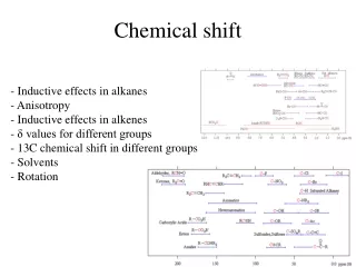
Water-Fat Signal Model • Single peak fat model • Multi peak fat model
Two-Point Dixon RF water water fat fat GPartition GPhase GReadout Op-phase In-phase Read out
Multi-Point Acquisition Op-phase 2 RF water water water fat fat fat GPartition GPhase GReadout Op-phase 1 In-phase Read out
Water-Fat Separation Methods • Image at echo time tl • Multi peak fat model
Water Fat Separation • Require the acquisition of two or more images at different TE • Long scan time needed • Compressed sensing can be combined with water-fat separation to improve sampling efficiency
Compressed Sensing • Key elements of a successful compressed sensing reconstruction: • Signal sparsity • Incoherent sampling • Nonlinear, sparsity promoting reconstruction
Nonlinear Reconstruction • Iterative reconstruction needed • Optimization based on minimizing l1 norm works well:
Imaging Parameters • 1.5 T scanner (Phillips Healthcare) • Retrospective under-sampling (Poisson-disk) • Knee images • Turbo spin echo, TR=500 ms, TE = 21 ms • FOV 160 mm x 160 mm • Matrix size 256 x 256, slice thickness 3mm, voxel size 0.6 mmx0.6 mmx3 mm • Echo time -0.4, 1.1, 2.6 ms (relative to spin echo)
Imaging Parameters • 1.5 T scanner (Phillips Healthcare) • Abdominal images • 3D gradient echo, TR=6.9 ms, TE1 = 1.66 ms, TE = 1.66 ms, =15 • FOV 400 mm x 320 mm x 216 mm • Matrix size 240 x 192 x 54, bandwidth 833 Hz/pix
Fat Signal Model • Single Peak Fat Model • Chemical shift of fat: -220 Hz • Multi Peak Fat Model • Three peak fat model: -30 Hz, -165 Hz, -210 Hz • Relative amplitude (0.15, 0.1, 0.75)
CS-WF Reconstruction • Initial field map estimation • Initialization: • Low-resolution: center k-space • High-resolution: perform CS reconstruction for each echo • Compute possible field map values for each pixel and estimate initial field map using region growing, and • Estimate initial water and fat images
CS-WF Reconstruction • Similar to Gauss-Newton algorithm • Iteratively and simultaneously update the water and fat images and the field map, using the update as:
CS-WF Reconstruction • Given the final estimate xn, compute a projection on k-space yn=g(xn), set the measured data at the sampling location yn=y|acq and perform one last iteration.
2D Knee Images Single peak fat model
2D Knee Images Error seems to have some texture
2D Knee Images Multi peak fat model (three peaks, three echoes)
CS-WF Reconstruction • Low-resolution initialization: 50 Gauss-Newton iterations • High-resolution initialization: 5 iteration • One Gauss-Newton step for 3D data: 9 min (This is slow!)
Discussion • Nice and uniform water fat separation • Good field map estimation • Clean image without noticable artifact • Slow reconstruction • Moderate reduction factor • High reduction factor results in loss of contrast
Study Based on This Paper • Silver HJ, et al. Comparison of gross body fat-water magnetic resonance imaging at 3 Tesla to dual-energy X-ray absorptiometry in obese women. Obesity (Silver Spring). 2013 Apr;21(4):765-74 • Pang Y, Zhang X. Interpolated compressed sensing for 2D multiple slice fast MR imaging. PLoS One. 2013; 8(2) • Pang Y, et al. Hepatic fat assessment using advanced Magnetic Resonance Imaging.Quant Imaging Med Surg. 2012 Sep;2(3):213-8 • Sharma SD, et al. Chemical shift encoded water-fat separation using parallel imaging and compressed sensing. MagnReson Med. 2013 Feb;69(2):456-66.
Study Based on This Paper • Li W, et al. Fast cardiac T1 mapping in mice using a model-based compressed sensing method. MagnReson Med. 2012 Oct;68(4):1127-34. • Sharma SD,et al. Accelerated water-fat imaging using restricted subspace field map estimation and compressed sensing. MagnReson Med. 2012 Mar;67(3):650-9