Download

1 / 60
800 likes | 1.48k Views
Bone Marrow Aspiration in Dogs and Cats. Wendy Blount, DVM. Practical Hematology. Determining the cause of anemia Treating regenerative anemias Blood loss Hemolysis Treating non-regenerative anemias Blood & plasma transfusions in general practice

E N D
Bone Marrow Aspiration in Dogs and Cats Wendy Blount, DVM
Practical Hematology • Determining the cause of anemia • Treating regenerative anemias • Blood loss • Hemolysis • Treating non-regenerative anemias • Blood & plasma transfusions in general practice • Determining the causing of coagulopathies • Treating coagulopathies in general practice • Finding the source of leukocytosis • Bone marrow sampling
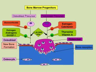
When Should You Do a Bone Marrow? BONE MARROW SAMPLING IS NO BIG DEAL • Any single cytopenia unresponsive to therapy • Bicytopenia or pancytopenia • Regenerative or nonregenerative? • Markedly high blood cell counts • Suspect leukemia
When Should You Do a Bone Marrow? • Looking for organisms that cause systemic infection • Histoplasma • Leishmania • Cytauxzoon • Looking for occult neoplasia • Hypercalcemia • Bony lysis on radiographs • Evaluating iron stores
When Should You NOT Do a Bone Marrow? • Severe coagulopathy • DIC • anti-vitamin K rodenticide toxicity • Severe liver failure • Severe anemia • Transfuse first prior to sedation
When Should You NOT Do a Bone Marrow? IS SEVERE THROMBOCYTOPENIA (<25,000/UL) A CONTRAINDICATION TO SAMPLING BONE MARROW? If secondary hemostasis is fine, NO • Fibrin, factors, fibrinolysis, vessels are OK • No DIC, no factor coagulopathy, no vasculitis, no problem • PT, PTT, BMBT normal – go ahead • Very few platelets are needed for effective primary hemostasis
Performing Bone Marrow Sampling Two procedures • Bone marrow aspirates • Bone marrow core biopsy Steps: • Preparing equipment • Patient preparation and sedation • Placement of the sampling needle • Procuring the sample • Preparing slides • Confirming adequate sampling • Submitting samples to the lab or interpreting results in house movie
Comments about theBone Marrow Aspiration Movie • There are a number of other positions for needle placement • Smaller volume of lidocaine (<3cc) may be adequate. • Careful of lidocaine in cats • I prefer to drape the area, to keep hands sterile • I prefer to coat the aspiration syringe with an anticoagulant • There is another technique for slide preparation which I think makes better slides
Choosing a Site Proximal humerus • There are 2 approaches • As shown in the video • As an IM pin would be placed. • Preferred for severe thrombocytopenia • Less soft tissue to go through • Direct pressure for primary hemostasis easily applied Proximal femur • Easier in the cat Wing of the ileum • Easier in the cat • 2 approaches – dorsal and lateral
Choosing a Needle Rosenthal needle • 100% metal (steel) Rosenthals can be re-autoclaved • May need to be sharpened occasionally • 16-18 gauge - not for biopsy
Choosing a Needle Illinois sternal-iliac Needle • guidepiece prevents deep penetration when penetrating wing of ileum • Take guidepiece off for long bones • 16-18 gauge – not for core biopsy • Can be re-autoclaved a few times or gas sterilized • Aluminum rather than steel – doesn’t sharpen well
Choosing a Needle Jamshidi needle • 13 to 8 gauge • For core biopsy or aspiration • Fine wire use to remove the core biopsy • End of the needle is tapered to retain the core
Choosing a Needle • Michel’s trephine • For lateral iliac wing marrow biopsies • Large dogs
Preparing Equipment Check your needle (sterile technique) • needle is sharp and without chips • stylet completely occludes the hollow needle • Stylet is properly seated in the needle
Preparing Equipment • Sterile supplies • Surgical gloves • keep paper wrap for sterile field • Drape • #11 blade • Inspected bone marrow needle • 10-12cc syringes • 18-gauge needles
Preparing Equipment • Non-sterile supplies • supplies for animal sedation • Surgical prep equipment • Lidocaine and syringes • EDTA or heparin • Petri dish or coffee mug • Pipettes • Microscope slides • Quick Stain (e.g., DiffQuick) • Microscope • Formalin and container if doing biopsy • Lab forms and mailers
Patient Preparation • Sedate • Clip and surgically prep site • 3-4 inches square • Lidocaine block down to bone, including periosteum • Re-scrub • drape
Needle Placement Proximal Humerus – “IM pin” method • Lateral recumbency • Rotate elbow medially and push humerus cranially to expose the shoulder • Stab incision #11 blade on “flat spot” between the greater tubercle and the humeral head • Thumb of other hand holding elbow along long axis of humerus for reference • Line needle up parallel with the long axis of the humerus (other thumb) • Make sure cap and stylet are in place • Slowly twist clockwise and counter-clockwise until needle is seated in cortical bone • Then screwdriver-like motion until needle well seated in the marrow cavity • Check needle by “wiggling” to make sure firmly seated in bone
Needle Placement Proximal Humerus – “IM pin” method
Needle Placement Proximal Humerus – “IM pin” method • Coat a 10-12cc syringe with anticoagulant • Have assistant attach sterile 18-gauge needle to syringe and remove cap • Assistant holds vial of anticoagulant • Draw up 1-1.5cc anticoagulant • Coat syringe by drawing plunger to highest cc mark on the syringe • Squirt anticoagulant into petri dish • Remove needle • Remove the cap and stylet from the bone marrow needle and place on the sterile field
Needle Placement Proximal Humerus – “IM pin” method • Firmly attach the coated syringe • Rapidly pull plunger back to 8-10cc • This hurts • As soon as you see blood, release pressure, and get 1cc or less of bone marrow • Very Quickly squirt the marrow into the petri dish, and swirl • Look for spicules (“flecks”) • If no spicules, remove needle and try again • If spicules, replace the marrow needle cap and prepare slides to confirm good sample • NOTE: leave needle in place if spicules
Needle Placement To Review – any location • Determine needle placement landmarks • Stab incision #11 needle • Line needle up at proper angle • Back and forth twisting until seated in cortex • Screwdriver-like motion until seated in marrow cavity • Can’t “feel” dropping into the marrow cavity • Coat syringe • Remove cap/stylet, attach syringe & aspirate • Squirt marrow in petri dish & look for flecks • If no flecks, remove needle and try again • If flecks, replace cap and prepare slides
Needle Placement - Landmarks Proximal Humerus – lateral technique • Lateral recumbency • Palpate the spine of the scapula, and then the acromion (a) • The next prominence is the greater tubercle (b) • Insert the needle at the distal end of the greater tubercle (c)
Needle Placement - Landmarks Proximal Humerus – lateral technique • The axis of the needle should be 45o to the long axis of the humerus
Needle Placement - Landmarks Proximal Humerus – lateral technique • The axis of the needle should be 45o to the long axis of the humerus ( ------- ) • Too far proximally will put you in the joint • Too much angle may put you in the bicipital bursa ( ------ ) • The needle has a tendency to slide distally as you are seating it in cortex, so seat the needle slowly and firmly prior to advancing into the cortex
Needle Placement - Landmarks Iliac Crest – dorsal technique
Needle Placement - Landmarks Iliac Crest – dorsal technique • Sternal recumbency, hindlimbs tucked under • Palpate “flat spot” on the iliac crest • Impossible in obese dogs • Easy in cats • Insert needle at widest point of the iliac crest • Axis of the needle is roughly perpendicular to the table • Direct slightly caudally rather than cranially • The needle has a tendency to slide off the iliac crest medially or laterally, so go slowly and firmly until seated in bone • Using the guidepiece of an Illinois needle can help control how far the needle penetrates if it slips off the crest
Needle Placement - Landmarks Iliac Crest – dorsal technique
Needle Placement - Landmarks Iliac Crest – dorsal technique
Needle Placement - Landmarks Iliac Wing – lateral technique
Needle Placement - Landmarks Iliac Wing – lateral technique • Not recommended for cats or small dogs (less than 25 lbs) • Lateral recumbency • Palpate “flat spot” on dorsal iliac crest • Insert the needle 1-2 cm ventral to the center of the iliac crest • Axis of the needle is perpendicular to the long axis of the ileum and perpendicular to the table • Careful not to advance the needle through the opposite cortex • Michels’ trephine or Jamshidi can be used to take a full thickness marrow/bone biopsy
Needle Placement - Landmarks Iliac Wing – lateral technique
Needle Placement - Landmarks Iliac Wing – lateral technique
Needle Placement - Landmarks Proximal femur • Easier in the cat (Jamshidi for large dogs) • Lateral recumbency • If right handed, sample the left rear leg • Rotate stifle slightly medially to open up the hip joint • Place non-dominant thumb along the long axis of femur, pointing proximally, and ending on the greater trochanter • Insert the needle under thumb, into the intertrochanteric fossa
Needle Placement - Landmarks Proximal femur • Once placed, move other hand down to the stifle, with thumb along long axis of femur • Twist back and forth until seated in cortex • Then hold knee with non-dominant hand to advance needle into the marrow cavity using a screwdriver- like motion
Needle Placement - Landmarks Proximal femur
Slide Preparation • Pipette flecks out of the petri dish and put on glass slides immediately • Elevate one end of the slide to let extra blood run off
Slide Preparation • Prepare gentle horizontal smears as well as vertical pull apart preps • Lymphoma cells are fragile and require vertical preps • Use a very light touch for the horizontal preps
Slide Preparation • Dry and stain a few slides to look for bone marrow cells • You’ll find the marrow in the clear spot on the unstained slides (displaces blood) • Dark spot on the stained slides
Slide Preparation • If you get blood with little marrow, either try another aspirate, or do a core biopsy • Keep unstained slides to submit with stained slide, in case special stains or restaining is needed • Work quickly, to prevent clotting • If clots form in the petri dish, you may be able to submit clotted samples in formalin for histopath • Difficult to confirm adequate sample on these
Aspiration or Core Biopsy? Advantages of aspiration • Cellular morphology is more clear • Better identification of cell lineages • Characteristics of malignancy • Can calculate E:M ratios • Estimate regenerative responses • Interpret with respect to CBC and reticulocyte count • Normal 3:1 to 5:1 • Maturation sequence counts are easier • More mature cell stages should be present in successively greater numbers • More younger cells means leukemia, maturation arrest or immune mediated destruction of the next stage
Aspiration or Core Biopsy? Advantages of core biopsy • If repeated attempts to aspirate produce no fluid (“packed marrow”) – myelophthisic disease • Myelofibrosis • (artifact) • If repeated attempts to aspirate produce blood only with flecks of fat • Aplastic anemia (hypocellular marrow) • Can evaluate marrow cellularity • Can evaluate tissue architecture • Invasion by normal looking lymphocytes indicates lymphoma • Can detect myelofibrosis or myelonecrosis
Bone Marrow Core Biopsy • Use Michel’s trephine – lateral iliac wing • Or Jamshidi in any approach • As soon as the needle is well seated through the cortex, remove the stylet, and replace the cap and handle • Advance the needle 1-2 cm further, rotating in a single direction • “stir” the needle to break loose the core • Remove the needle rotating in a single direction • Pass the wire or stylet backward to pop the core out the top of the needle • Core 0.75-1 cm long is sufficient • Cytologies can be made by rolling the core on slides, or scraping it • Place cores in formalin for histopathology
Evaluation of Adequate Sampling Gross examination • Unstained slide – blank spot • Stained slide – very dark purple spot • Bone marrow may require longer staining Microscopic examination • Large nucleated cells confirm presence of bone marrow • Blue round cells erythroid • “pink squigglies” are myeloid
Evaluation of Adequate Sampling • Peripheral blood only indicates poor sample • May see dark brown iron stores at low power