Download
1 / 10
170 likes | 528 Views
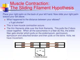
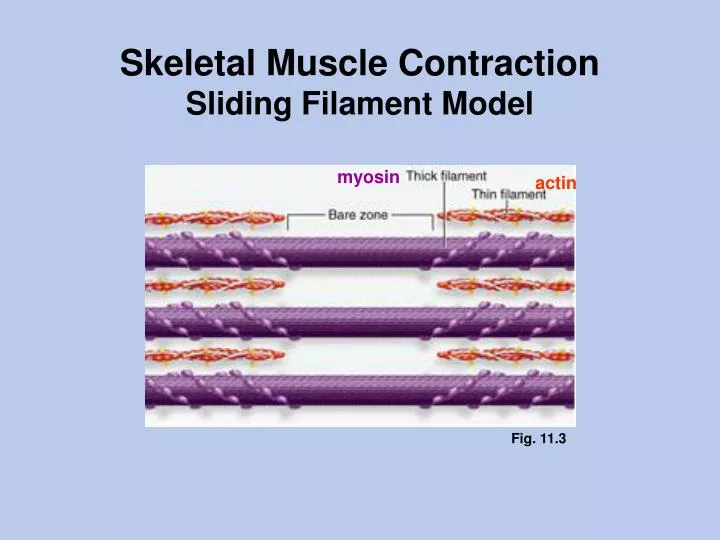
Skeletal Muscle Contraction Sliding Filament Model. myosin. actin. Fig. 11.3. Thin (actin) filament. Fig. 3-3 Ganong. . . . . . . . . . actin monomer G-actin (globular actin). actin polymer F-actin (filamentous actin). from Alberts et al., Molecular Biology

E N D
Skeletal Muscle ContractionSliding Filament Model myosin actin Fig. 11.3
Thin (actin) filament Fig. 3-3 Ganong actin monomer G-actin (globular actin) actin polymer F-actin (filamentous actin) from Alberts et al., Molecular Biology of the Cell Fig. 11.3
Thick (myosin) filament Fig. 11.3 myosin molecule (“monomer”): 2 heavy chains + 4 light chains central bare zone from Alberts et al., Molecular Biology of the Cell
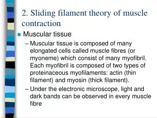
Striated Muscle Fig. 11.1 Fig. 3-2 Ganong A- band (anisotropic) contains thick filaments (and portions of thin filaments) I- band (isotropic) contains thin filaments Fig. 3-3 Ganong
Striated Muscle Fig. 11.4
Striated Muscle Fig. 11.2
Sliding Filament Model of Contraction Fig. 11.9 Fig. 3.3 Ganong
Fig. 11.9 Cross Bridge Cycle The myosin head is an ATPase. The two most important shape-changing events are 13/10 ATP binding (which leads to detachment and reorientation) 11/12 Pi release (which leads to the power stroke) causes Pi to be released. As myosin heads bind ATP, the crossbridges detach from actin, become reoriented and hydrolyze ATP to ADP and Pi. Pi No ATP no detachment e.g., rigor mortis Power stroke causes ADP to be released
ATP binding to myosin ATP binding is more important for reorientation than ATP hydrolysis. Fig. 3-6 Ganong (19th edition) The image above and the modifications to the Saladin text in the previous slide are based on Raiment et al., Science 261:50-58, 1993, and Vale and Milligan, Science 288:88-95, 2000. (see also Fig. 16.58 in Alberts et al., Molecular Biology of the Cell, 4th ed., 2002)
Length-Tension Relationship Fig. 11.11 ly short Increased muscle diameter causes increased separation (the lattice spacing) between thick and thin filaments. (actual mechanism still a topic of debate, see Fuchs and Martyn, Length-dependent Ca2+ activation in cardiac muscle: some remaining questions. J. Muscle Res. and Cell Motility, 26:199-212, 2005) Increased muscle length causes decreased overlap between thick and thin filaments. = normal operating length for cardiac muscle = normal operating length for skeletal muscle