Download
1 / 22
220 likes | 242 Views
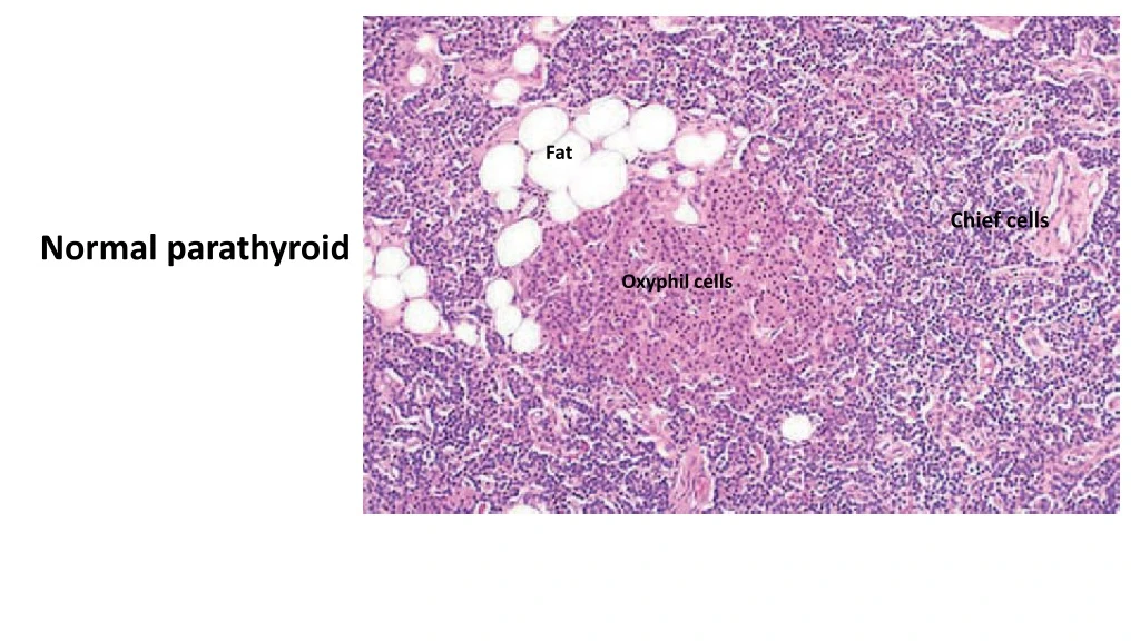
Fat. Chief cells. Normal parathyroid. Oxyphil cells. Parathyroid adenoma. capsule. Normal parathyroid. Incidental cyst (not important). Tumor behavior is much more important than morphology. The neoplasm. Parathyroid carcinoma. Tumor invasion in the capsule.

E N D
Fat Chief cells Normal parathyroid Oxyphil cells
Parathyroid adenoma capsule Normal parathyroid Incidental cyst (not important) Tumor behavior is much more important than morphology The neoplasm
Parathyroid carcinoma Tumor invasion in the capsule
Parathyroid hyperplasia Same as normal tissue but expanded …and somehow, adipose tissue is diminished
Islet of Langerhans Normal pancreas Exocrine pancreas (acini with zymogenic color of the cells)
Inflamed islet Insulitis DM type 1 Lymphocytes
Amyloid Advanced stage of DM2 Islet of Langerhans
Trabeculae of tumor cells …they can grow in many patterns …here: trabecular pattern Islet cell tumor (= pancreatic neuroendocrine tumor) Normal pancreas Islet May be benign May be malignant The tumor behavior is the most important
Islet cell tumor also a trabecular pattern is seen here …resembling trabecular bone This is a neuroendocrine tumor (like carcinoid) So when you look at the nuclei:
Cortex Normal adrenal gland Zona glomerulosa Medulla Zona reticularis Zona fasciculata Cells with good amount of basophilic cytoplasm (chromaffin cells)
Atrophic adrenals e.g., exogenous steroids (low ACTH) or Addison Golden cortex Normal adrenals Dark medulla Hypertrophic glands e.g., ACTH oversecretion
Neuroblastoma Neuroblastoma that have grown to the degree of displacing the liver to the left Liver
An adrenal medullary tumor …mainly in children
*It is a small round blue cell tumor *High mitotic and apoptotic activity and necrosis mitoses
Adrenocortical adenoma A well-circumscribed adrenocortical tumor
Adrenocortical adenoma Resembling adrenocortical cells
Adrenocortical carcinoma Discovered at an advanced stage because the retroperitoneal space permits this degree of enlargement
Pheochromocytoma An adrenal medullary tumor After staining with dichromate...due to the oxidation of catecholamines
Pheochromocytoma May be benign May be malignant The most important is behavior Part of pheochromocytoma tumor …the cells have good amount of basophilic cytoplasm Cortex
Capillary network Nests of polygonal to spindle-shaped chromaffin cells (zellballen pattern) that are supplied by a rich vascular network Pheochromocytoma
Pheochromocytoma Capillary network Nest This nested pattern is called: Zellballen pattern Nest Nest Sustentacular cells