Download
1 / 35
350 likes | 516 Views
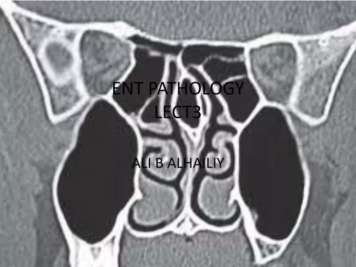
ENT PATHOLOGY LECT3. ALI B ALHAILIY. ENT (ear, nose, and throat) is the branch of medicine and surgery that specializes in the diagnosis and treatment of disorders of the head and neck. NASAL CAVITY. Indications.

E N D
ENT PATHOLOGYLECT3 ALI B ALHAILIY
ENT (ear, nose, and throat) is the branch of medicine and surgery that specializes in the diagnosis and treatment of disorders of the head and neck.
Indications > Visual loss > Vascular lesions of the orbit (AV malformation , carotid-cavernous fistula, orbital varix)> History of visual disturbances > Optic nerve sheath meningioma> Tumours or suspected tumours> Thyroid ophtalmopathy> Extraocularmyopathy> Optic nerve neuritis> Optic nerve glioma> Orbital abscess> Inflammation
1- SINUSITIS • Sinusitis is inflammation of the paranasal sinuses. It can be due to infection, allergy, or autoimmune problems. • Most cases are due to a viral infection and resolve over the course of 10 days. • It is a common condition, with over 24 million cases annually in the U.S
Signs and symptoms • Headache • Facial pain • constant, or aching sort over the affected sinuses is common with both acute and chronic stages of sinusitis • Sinus infections can also cause inner ear problems due to the congestion of the nasal passages
DIAGNOSIS • For sinusitis lasting more than 12 weeks a CT scanis recommended. • Nasal endoscopy, and clinical symptoms are also used to make a positive diagnosis. • A tissue sample for histology andculturescan also be collected and tested
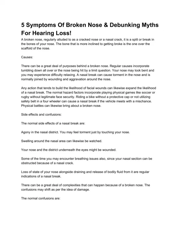
2- Facial Trauma • Facial trauma, also called maxillofacial trauma, is any physical trauma to the face. • Facial trauma can involve soft tissue injuries such as burns, lacerations and bruises, or fractures of the facial bones such as nasal fractures and fractures of the jaw, as well as trauma such as eye injuries
Sign and Symptoms • Symptoms are specific to the type of injury; for example, fractures may involve pain, swelling, loss of function, or changes in the shape of facial structures. • Facial injuries have the potential to cause loss of function; for example, blindness or difficulty moving the jaw can result. • Although it is seldom life-threatening, facial trauma can also be deadly, because it can cause severe bleeding or interference with the airway
Fractures of facial bones, like other fractures, may be associated with pain, bruising, and swelling of the surrounding. • Fractures of the nose, base of the skull, or maxilla may be associated with nosebleeds. • Nasal fractures may be associated with deformity of the nose, as well as swelling and bruising. • Deformity in the face, for example a teeth which do not align properly, suggests the presence of fractures. • People with mandibular fractures often have pain and difficulty opening their mouths and may have numbness in the lip and chin.
Diagnosis • Radiological imaging of tissues using X-rays, is used to rule out facial fractures .However the complex bones and tissues of the face can make it difficult to interpret plain radiographs. • CT scanning is better for detecting fractures and examining soft tissues, and is often needed to determine whether surgery is necessary. • CT scanning is usually considered to be more definitive and better at detecting facial injuries than X-ray. • CT scanning is especially likely to be used in people with multiple injuries who need CT scans to assess for other injuries anyway.
Thyroid cancer • Thyroid cancer is a malignant neoplasm originating from follicular or parafollicularthyroid cells. • Thyroid epithelial cells (also called follicular cells or principal cells) are cells in the thyroid gland that are responsible for the production and secretion of thyroid hormones, that is, thyroxine (T4) and triiodothyronine (T3). • Parafollicular cells (also called C cells) are neuroendocrincells in the thyroid with primary function to secrete calcitonin (It acts to reduce blood calcium (Ca2+), opposing the effects of parathyroid hormone (PTH ). • They are located adjacent to the thyroid follicles and reside in the connective tissue.
Classification • Thyroid cancers can be classified according to their histopathological characteristics • Papillary thyroid cancer (75% to 85% of cases ) – often in young females – excellent prognosis. • Follicular thyroid cancer (10% to 20% of cases ). • Medullary thyroid cancer (5% to 8% of cases)- cancer of the parafollicularcells.
Signs and symptoms • Most often the first symptom of thyroid cancer is a nodule in the thyroid region of the neck.However, many adults have small nodules in their thyroids, but typically under 5% of these nodules are found to be malignant. • Sometimes the first sign is an enlarged lymph node. Later symptoms that can be present are pain in the anterior region of the neck . • changes in voice due to an involvement of the recurrent laryngeal nerve. • Thyroid cancer is usually found in a euthyroidpatient, but symptoms of hyperthyroidism or hypothyroidism may be associated with a large or metastatic well-differentiated tumor. • Thyroid nodules are of particular concern when they are found in those under the age of 20. The presentation of benign nodules at this age is less likely, and thus the potential for malignancy is far greater.
Diagnosis • After a thyroid nodule is found during a physical examination. • Most commonly an ultrasound is performed to confirm the presence of a nodule, and assess the status of the whole gland. • Measurement of thyroid stimulating hormone and anti-thyroid antibodies will help decide if there is a functional thyroid disease present, a known cause of a benign nodular goiter. • Measurement of calcitonin is necessary to exclude the presence of medullary thyroid cancer. • Finally, to achieve a definitive diagnosis before deciding on treatment, a fine needle aspiration cytology test is usually performed. • CT scan can be performed to see any metastatic lesions
Treatment • The most effective management of aggressive thyroid cancers is • 1.surgical removal of thyroid gland (thyroidectomy) followed by 2. radioactive iodine ablation and TSH-suppression therapy. 3.Chemotherapy or radiotherapy may also be used in cases of distant metastases or advanced cancer stage.
Axial CT Scan With IV contrast Without IV contrast
AXIAL CORONAL
Papillary mass MRI AXIAL MRI CORONAL