Download
1 / 55
550 likes | 758 Views
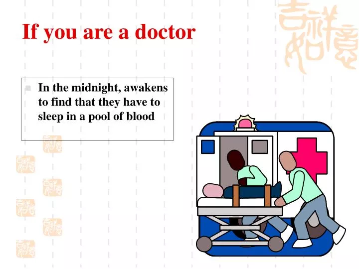
If you are a doctor. In the midnight, awakens to find that they have to sleep in a pool of blood. How to diagnosis ? How to management ?. You. Antepartum Hemorrhage. Obstetrics & Gynecology Hospital of Fudan University Xu Huan. Rationale (why we care…).

E N D
If you are a doctor • In the midnight, awakens to find that they have to sleep in a pool of blood
How to diagnosis? How to management? You
Antepartum Hemorrhage Obstetrics & Gynecology Hospital of Fudan University Xu Huan
Rationale (why we care…) • 4-5% of pregnancies complicated by 3rd trimester bleeding • Immediate evaluation needed • Significant threat to mother & fetus (consider physiologic increase in uterine blood flow) • Consider causes of maternal & fetal death • Priorities in management (triage!)
Objectives • We will be able to: • Describe the approach to the patient with third-trimester bleeding • Compare symptoms, physical findings, and diagnostic methods that differentiate bleeding etiologies • Describe management and delivery options for 3rd trimester bleeding etiologies • Describe potential maternal and fetal morbidity & mortality • Describe management of postpartum hemorrhage • Apply knowledge in the discussion of clinical case scenarios
Vaginal Bleeding: Differential diagnosis • Common: • Abruption, previa, preterm labor, labor • Less common: • Uterine rupture, fetal vessel rupture, lacerations/lesions, cervical ectropion, polyps, vasa previa, bleeding disorders • Unknown • NOT vaginal bleeding!!! (happens more than you think!)
Other Etiologies • Cervicitis • infection • Cervical erosion • Trauma • Cervical cancer • Foreign body • Bloody show/labor
Perinatal mortality and morbidity • Previa • Decreased mortality from 30% to 1% over last 60 years • Now emergent cesarean delivery often possible • Risk of preterm delivery • Abruption • Perinatal mortality rate 35% • Accounts for 15% of 3rd trimester stillbirths • Risk of preterm delivery • Most common cause of DIC in pregnancy • Massive hemorrhage --> risk of ARF, Sheehan’s, etc.
Definition • After 28 pregnant weeks placental implantation over the cervical os or in the lower uterine segment • It constitutes an obstruction of descent of the presenting part • Main cause of obstetrical hemorrhage(20%) • Incidence 0.24%-1.57% (our country).
Risk factors & Associations • Prior cesarean delivery/myomectomy • Prior previa (4-8% recurrence risk) • Previous abortion • Increased parity • Multiple pregnancy • Advanced maternal age • Abnormal presentation • Smoking
Etiology • Causes • Endometrial abnormality • Scared or poorly vascularized endometrium in the corpus. • Curettage, Delivery, CS and infection of endometrium • Placental abnormality Large placenta (multiple pregnancy), succenturiate lobe • Delayed development of trophoblast
Classification Complete placenta previa Partrial placenta previa Marginal placenta previa
Manifestation(1) Symptoms • Painless vaginal bleeding (70%) • Spontaneous,After coitus • The most characteristic symptom • late pregnancy (after the 28th week) and delivery • Characteristics: sudden, painless and profuse • Contractions • No symptoms • Routine ultrasound finding • The mean gestational age of first bleed: 30 wks • 1/3 before 30 weeks
Manifestation(2) • Anemia or shock repeated bleeding→ anemia heavy bleeding→ shock • Abnormal fetal position a high presenting part breech presentation (often)
Physical Findings • Bleeding on speculum exam • Cervical dilation • Bleeding a sx related to PTL/normal labor • Abnormal position/lie • Non-reassuring fetal status • If significant bleeding: • Tachycardia • Postural hypertension • Shock
Diagnosis(1) • History • Painless hemorrhage • At late pregnancy or delivery • History of curettage or CS
Diagnosis(1) • Signs • Abdominal findings • Uterus is soft, relaxed and nontender. • Contraction may be palpated. • A high presenting part can’t be pressed into the pelvic inlet. Breech presentation • Fetal heart tones maybe disappear (shock or abruption)
Diagnosis • Speculum examination Rule out local causes of bleeding, such as cervical erosion or polyp or cancer. • Limited vaginal examination (seldom used) Palpation of the vaginal fornices to learn if there is an intervening bogginess between the fornix and presenting part. • Rectal examination is useless and dangerous
Diagnosis(1) • Ultrasound • abdominal 95% accurate to detect • transvaginal (TVUS) will detect almost all • consider what placental location a TVUS may find that was missed on abdominal • MRI • Check the placenta and membrane after delivery • remember: no digital exams unless previa RULED OUT!
Diagnosis • Before 20 weeks’ gestation,4-6% have some degree of placenta previa on ultrasonic examination • 90% of these resolving by the third trimester • Only 10% of complete placenta
Differential Diagnosis • Placental abruption vagina bleeding with pain, tenderness of uterus. • vasa previa In cases of velamentous cord insertion fetal vessels cover cervical os • Abnormality of cervix cervical erosion or polyp or cancer
Effects • obstetrical hemorrhage • Placenta accreta, increta, and percreta • Anemia and infection • Premature labor or fetal death or fetal distress
Treatments • Expectant therapy • Rest: keep the bed • Controlling the contraction: MgSO4 • Treatment of anemia • Preventing infection
Treatments • Termination of pregnancy • CS • total placenta previa (36th week), Partial placenta previa (37th week) and heavy bleeding with shock • Preventing postpartum hemorrhage: pitocin and PG • Hysterectomy: Placenta accreta or uncontroled bleeding
Treatments • Vaginal delivery Marginal placenta previa Vaginal bleeding is limited
Management • Initial evaluation/diagnosis • Observe/admit to L&D • IV access, routine (maybe serial) labs • Continuous electronic fetal monitoring • Continuous at least initally • May re-evaluate later if stable, no further bleeding • Delivery???
Management • Less than 36 wks gestation -expectant management if stable, reassuring • Bed rest (negotiable) • No vaginal exams (not negotiable) • Steroids for lung maturation (<32 wks) • Possible mgmt at home after 1st bleed • 70% will have recurrent vaginal bleeding before 36 completed weeks requiring emergent cesarean
Management • 36+ weeks gestation • Cesarean delivery if positive fetal lung maturity by amniocentesis • Delivery vs expectant mgmt if fetal lung immaturity • Schedule cesarean delivery at 37 weeks • Discussion/counseling regarding cesarean hysterectomy • Note: given stable maternal and reassuring fetal status, none of these management guidelines are absolute (this is why Obstetrics is so much fun!)
Other Considerations • Placenta accreta, increta, percreta • Cesarean delivery may be necessary • History of uterine surgery increases risk • Must consider these diagnoses if previa present • Could require further evaluation, imaging (MRI considered now) • NOT the delivery you want to do at 2 am
A B Abnormally adherent placentation. A. Placenta accreta. B. Placenta increta. C. Placenta percreta C
Definition • abruptio placentae or placental abruption: placental separation from its implantation site before delivery (the normally implanted placenta ) • Incidence • complicates 0.5-1.5% of all pregnancies • recurrence risk • 10% after 1st episode • 25% after 2nd episode
Cocaine maternal hypertension abdominal trauma smoking prior abruption preeclampsia multiple gestation prolonged PROM uterine decompression short umbilical cord chorioamnionitis multiparity Risk factors & Associations
Pathology • Placental separation is initiated by hemorrhage into the decidua basalis with formation of a decidual hematoma • Concealed hemorrhage • Revealed hemorrhage
revealed hemorrhage concealed hemorrhage
Total placental abruption with concealed hemorrhage and fetal death
Maternal-fetal risk • perinatal mortality: 35% • DIC • hypovolemic shock • acute renal failure • Sheehan’s syndrome
Symptoms • Vaginal bleeding • Abdominal or back pain • Uterine contractions • Uterine tenderness
Physical Findings • Vaginal bleeding • Uterine contractions • Hypertonus • Tetanic contractions • Non-reassuring fetal status or demise • Can be concealed hemorrhage
Laboratory Findings • Anemia • may be out of proportion to observed blood loss • DIC • Can occur in up to 10% (30% if “severe”) • First, increase in fibrin split products • Followed by decrease in fibrinogen
Diagnosis • Clinical scenario • Physical exam • Not digital pelvic exams until rule out previa • Careful speculum exam • Ultrasound • Can evaluate previa • Not accurate to diagnose abruption