Download
1 / 28
280 likes | 531 Views
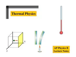
Diffusion Physics - Thermal Agitation. In steady state, the motion of water is dominated by thermal agitation. This causes “random” motion of water within a compartment. Diffusion Physics - Thermal Agitation. The “rate” of water motion is determine by a diffusion coefficient, “D”.

E N D
Diffusion Physics - Thermal Agitation • In steady state, the motion of water is dominated by thermal agitation. • This causes “random” motion of water within a compartment.
Diffusion Physics - Thermal Agitation • The “rate” of water motion is determine by a diffusion coefficient, “D”. • Mean displacement of water molecules is related to “D” by Einstein’s equation:
Detecting Diffusion with MRI - Intravoxel Incoherent Motion From Ellingson, Concepts in MR, 2008
Detecting Diffusion with MRI - Intravoxel Incoherent Motion Phase of “Tagged” H2O Function of Diffusion Gradients Detected DWI Signal Diffusion Coefficient MRI Signal w/o Diffusion Sensitivity
Factors that affect diffusion coefficient, D • Diffusion Time, t • Physical time between gradients used to “tag” H2O • Size of Compartment(s) • If we set a limit for r, then we observe an apparent diffusion coefficient, ADC • Tortuosity of the Compartments • - More tortuous paths look like slow diffusion • Temperature • Viscosity *** We can only measure “ADC” because of all the factors that change “D”! ***
Diffusion Tensor Imaging (DTI) • Diffusion anisotropy can occur when compartments are not symmetric • Diffusion may be higher in different directions • To determine diffusion anisotropy, we use Diffusion Tensor Imaging (DTI) • DTI uses diffusion sensitizing gradients to determine ADC in different directions • From these measurements we construct the mathematical tensor • In this way, DTI more accurately models the geometry of tissues Isotropic Diffusion Anisotropic Diffusion
Diffusion Tensor Imaging (DTI) The Diffusion Tensor: Isotropic Diffusion 1 = 2 = 3 Anisotropic Diffusion 1 > 2,3 From Ellingson, Concepts in MR, 2008
Diffusion MR Characteristics of theCentral Nervous System • MR constraints largely limit ADC measurement to the extracellular compartment. • Diffusion in the spinal cord is highly anisotropic for both gray matter and white matter Davidoff, A., Handbook of the Spinal Cord From Ellingson, Concepts in MR, 2008 From A.Todd, Univ. Glascow
Diffusion MR Characteristics of theCentral Nervous System • Transverse ADC (tADC) is dependent on (Schwartz, 2005) • Axon Counts = tADC • Extracellular Volume = tADC • Myelin Volume = tADC • Longitudinal ADC (lADC) is dependent on (Song, 2003; 2002; Sun, 2006; Ellingson, 2008) • Structural and Functional Integrity of Axons • Microfilament & Neurofilament Density • Axonal Transport System Integrity From Ellingson, Concepts in MR, 2008
DTI Tractography • In the CNS, lADC > tADC & 1 is parallel to axon orientation
Diffusion MR Characteristics of theCentral Nervous System • Anisotropy is preserved across surrogates (Ellingson, Concepts in MR, 2008) • ADC is consistent across pulse sequences (Ellingson, AJNR, 2008) Ellingson, 2008
Diffusion MR Characteristics of theCentral Nervous System Human • ADC changes across the length of the spinal cord (Ellingson, AJNR, 2008; Ellingson, JMRI, 2008) Rat Rostral Caudal
Mechanical Injury: Stretching and tearing of axons immediate death of all damaged cells. Hypoxia and Ischemia: Blood flow to injury is restricted. Anaerobic metabolic processes in viable tissue, other tissues become necrotic. Hemorrhage & Vasogenic Edema: In contusion injury, hemorrhages start in central gray matter and spread radially. Early vasogenic edema forms due to blood constituents leaving vasculature. Damage to Axon Transport Systems: Microtubules and neurofilaments begin to degrade. Pathology of Acute SCI Normal Mechanical Injury Vasogenic Edema From Ellingson, Concepts in MR, 2008
Mechanical Injury: Overall ADC at lesion site due to lack of boundaries to diffusion Hypoxia and Ischemia: Cause ADC DTI in Acute SCI • Hemorrhage & Edema: • Causes ADC at lesion site due to lack of boundaries to diffusion • Damage to Axon Transport Systems: • Causes lADC From Ellingson, Concepts in MR, 2008 lADC tADC Rat - Ex Vivo (Ellingson, JMRI, 2008b)
Reactive Cells: Active microglia increase in density Active Astrocytes hypertropy and line the cavity wall Anterograde and Retrograde Degeneration: Axons form retraction bulbs at proximal ends Demyelination occurs Proximal axons begin to swell Distal axons are dissolved Pathology of Subacute SCI • Gray Matter Morphological Changes: • Damaged axon nuclei move to eccentric locations within the cell body. • Neurons begin to hypotrophy and dendrites retract • Cytotoxic Edema • Axons and soma swell, the extracellular space decreases Reactive Cells Normal Cytotoxic Edema From Ellingson, Concepts in MR, 2008
Reactive Cells: Glial scar changes 1 orientation (Schwartz, 2005) High density microglia ADC Tortuosity from activated astrocytes causes ADC Anterograde and Retrograde Degeneration: tADC during demyelination lADC due to axon transport damage ADC due to axon swelling DTI in Subacute SCI • Gray Matter Morphological Changes: • Vasogenic Edema • Axons and soma swell, the extracellular space decreases • This causes ADC From Ellingson, Concepts in MR, 2008 Rat - Ex Vivo (Ellingson, JMRI, 2008b) tADC lADC
Long-term Axon Degeneration Progressive demyelination Loss of large diameter axons Widespread spreading of cysts Retrograde & Transneuronal degeneration damages whole spinal tracts Pathology of Chronic SCI • Gray Matter Morphology Changes • Chromatolysis • Nuclear Changes • Spinal Cord Atrophy Normal Atrophy + Axon Loss Normal MNs Chronic SCI MNs From Ellingson, Concepts in MR, 2008
DTI in Chronic SCI Rat - Ex Vivo (Ellingson, JMRI, 2008b) Remote Changes Cavity Formation
DTI in Chronic SCI Rat - Ex Vivo (Ellingson, JMRI, 2008b)
DTI in Chronic SCI Chronic Human SCI (Ellingson, AJNR, 2008b) Rostral-Caudal Asymmetry at Lesion Epicenter C5 Complete Injury
Functional Correlates of DTI Axonal Damage (Song, 2003; 2002; Nair, 2005; Sun, 2006) ↓ Longitudinal ADC (lADC) Myelin Damage (Song, 2003; 2002; Nair, 2005; Sun, 2006) ↑ Transverse ADC (tADC)
Normal SCI No temporal Coherence Loss of Amplitude Functional Correlates of DTI
Ad-fiber input to MSTT (Valeriani, 2007; Latash, 1988) C-fiber input to LSTT (Valeriani, 2007; Li, 1991; Latash, 1988) Functional Correlates of DTI Ellingson, J Neurotrauma, 2008, In Press
Functional Correlates of DTI From Ellingson, Biomed Sci Instrum, 2008 & Congress Neurological Surgeons, 2008
DTI is highly sensitive to the structural integrity of the spinal cord. DTI can be used to monitor the progression of SCI from acute through chronic stages DTI may also be sensitive to the functional integrity of the spinal cord. Future studies will be aimed at determining if DTI can predict long-term functional recovery in incomplete SCI Conclusions
Brian Schmit, Ph.D., Dept. of Biomedical Eng., Marquette University Shekar Kurpad, M.D., Ph.D., Dept. of Neurosurgery, MCW John Ulmer, M.D., Dept. of Radiology, MCW Maria Crowe, Ph.D., Dept. of Neurosurgery, MCW Kathleen Schmainda, Ph.D., Dept. Radiology & Biophysics, MCW Kristina Ropella, Ph.D., Dept. Biomed. Eng., Marquette University Funding: NIH R21, Brian Schmit (PI) Falk Foundation Marquette University VA Medical Center, Milwaukee, WI Bryon Riesch Paralysis Foundation Thank You