Download
1 / 48
510 likes | 669 Views
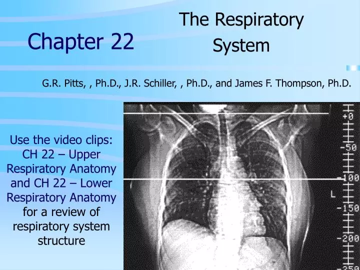
Chapter 22. The Respiratory System. G.R. Pitts, , Ph.D., J.R. Schiller, , Ph.D., and James F. Thompson, Ph.D. Use the video clips: CH 22 – Upper Respiratory Anatomy and CH 22 – Lower Respiratory Anatomy for a review of respiratory system structure. Respiration. Pulmonary ventilation

E N D
Chapter 22 The Respiratory System G.R. Pitts, , Ph.D., J.R. Schiller, , Ph.D., and James F. Thompson, Ph.D. Use the video clips: CH 22 – Upper Respiratory Anatomy and CH 22 – Lower Respiratory Anatomy for a review of respiratory system structure
Respiration • Pulmonary ventilation • Breathing - inspiration & expiration • External (pulmonary) respiration • Gas exchange between lung (alveoli) & blood • Transport of respiratory gases • Oxygen and carbon dioxide (CO2) must be transported between the tissues and the lungs • Internal (tissue) respiration • Gas exchange between blood and tissue cells • RBCs deliver O2 and pick up CO2 in the capillary beds • cells use O2 and produce CO2 (cellular respiration)
The Respiratory Tree • upper respiratory tract for ventilation (conduction of air) • lower respiratory tract for respiration (gas exchange by diffusion)
The Larynx Voice Production • Vestibular folds (false vocal cords) • Vocal folds (true vocal cords) • during exhalation laryngeal muscles pull the folds across the opening and tense the folds • exhaled air induces vibrations which create sound waves • volume • pitch
Regulation of the Airway • Smooth muscle • Parasympathetic (ANS), allergic response - bronchoconstriction • Sympathetic (ANS) response - bronchodilation • histamine release • allergy/asthma
The Alveolar Space • Alveolar fluid • Surface tension • Attraction of water to other water molecules • Surfactant: phospholipids decrease surface tension • Respiratory distress syndrome
The Pleural Cavities • Lungs – housed in the bony thorax • Pleural problems • pneumothorax • hemothorax • pleurisy: inflammation • collapsed lung: atelectasis
Muscles For Ventilation • Muscles for inspiration • diaphragm • dome-shaped muscle forms inferior wall of thoracic cavity • muscle flattens when contracted, expanding thoracic cavity • minimal involvement in normal resting breathing • important for physical exertion and speech/singing • can be limited by tight clothing, pregnancy, obesity, edema • external intercostals • pull ribs upward, push sternum forward, expand thoracic cavity
Muscles For Ventilation • Muscles for expiration • internal intercostals • pull ribs downward, pull sternum inward, compress thoracic cavity • abdominals • compress abdominal and thoracic cavities
Pulmonary Ventilation • Exchange of gases between the atmosphere and the alveoli of the lung • Bulk flow of gases due to pressure differences • Lung air pressure is atmospheric (760 mm Hg at sea level) • need to create a pressure gradient for air flow into the lungs (Q=ΔP/R) • two mechanisms • increase atmospheric pressure (positive ventilation) • decrease lung air pressure (negative ventilation) • Structure & Function of thoracic cavity helps
Physics of Ventilation • Boyle’s law - pressure in a closed container is inverselyproportional to the volume of container • Diaphragm, pleura and thoracic wall • At rest, volume decreased • During inspiration, volume increased
Ventilation Pressure Relationships • Intrapulmonary pressure (Ppul) • In alveoli • Variable, but equilibrates with atmospheric (760 mm Hg at sea level) • Intrapleural pressure (Pip) • Pleural cavity • Usually 4 mm Hg less that Ppul • Lungs have elastic recoil • Pleural fluid surface tension • Elasticity of chest wall • Transpulmonary pressure = (Ppul - Pip)
Pul. Ventilation - Inspiration • Pressure changes • With expansion of the rib cage and depression of the diaphragm, intrapulmonary pressure falls 1-2 mm Hg • Establishes a small negative pressure gradient permitting air flow into lungs
Pulmonary Ventilation - Expiration • Breathing out (expiration) also due to a pressure gradient • 3 important factors: • relaxation of diaphragm (rises) • elastic recoil of chest wall and the lungs • surface tension the pleural and alveolar fluids of the lungs • Forced muscular expiration – oblique and transverse abdominals indirectly “compress" the lungs
Ventilation Assessment • Respiration (ventilation) • 1 ventilatory cycle (1 inspiration and 1 expiration) • 12 breaths/min (resting rate = RR) • minute ventilation - ~6 L/min • Pulmonary Volumes & Capacities • Spirometry: measures respiratory volumes on a spirogram (recording) • [Biopac exercises in lab]
Pulmonary Volumes (measured) • Tidal volume (TV) • 350 mL reaches the alveoli • 150 mL do not, this air is trapped in anatomical dead space • Inspiratory reserve volume (IRV) • Expiratory reserve volume (ERV) • Residual volume (RV) [~1 L] • FEV1 – forced expiratory volume in 1 second
Pulmonary Capacities (calculated) • Pulmonary capacities = sums of certain lung volumes • Inspiratory capacity (IC) = TV+IRV [~ 3600 mL] • Functional residual capacity (FRC) = ERV+RV • Vital capacity (VC) = IRV+TV+ERV [~4800 mL] • Total lung capacity (TLC) = sum of all volumes
Exchange of O2 & CO2 - Gas Laws • Dalton's law • Each gas in a mixture of gases exerts own pressure as if all other gases were not present • Atmospheric pressure = sum of all partial pressures (p) of atmospheric gases • atmospheric pressure at sea level - 760 mm Hg • N2 - 79% - 600 mm Hg • O2 - 21% - 160 mm Hg, 105 mm Hg in alveoli • CO2 - 0.04% - 0.30 mm Hg • partial pressure difference with increasing altitude • 10,000 ft - 523 mm Hg - pO2 110 mm Hg (67 mm Hg in alveoli) • 20,000 ft - 349 mm Hg - pO2 = 73 mm Hg (40 mm Hg in alveoli) • 50,000 ft - 87 mm Hg, pO2 = 18 mm Hg (2 mm Hg in alveoli) • partial pressure difference with diving depth under water • 33 ft - 1520 mm Hg - pO2 320 mm Hg (210 mm Hg in alveoli) • pressure increases 1 atmosphere for every 33 ft of increased depth
Exchange of O2 & CO2 - Gas Laws • Henry's law • Amount of a gas that dissolves in liquid is proportional to the partial pressure of gas and its solubility coefficient • Solubility coefficients for normal gases • O2 • 0.024 ml O2 /mm Hg • 2.5 ml O2 at atmospheric pressure • CO2 • 0.57 ml/mm Hg • high solubility, low % • N2 • 0.012 ml/mm Hg • low solubility, high % • Nitrogen narcosis • Bends
Exchange of O2 and CO2 • Gas exchange between alveoli & capillaries = external respiration • changing deoxygenated to oxygenated blood • rate of gas exchange is dependent on: • surface area for diffusion • diffusion distance • pressure gradient • breathing rate/depth
Exchange of O2 and CO2 (cont.) • Internal (tissue) respiration • O2 & CO2 exchange between capillaries and tissue cells • changing oxygenated to deoxygenated • Only 25% of the blood’s O2 enters the cells at rest • CO2 moves in the opposite direction • Diffusion is driven by pressure gradients (and concentration gradients)
O2 Transport In The Blood • O2 does not dissolve well in water • another mechanism is needed to carry O2 • most O2 is carried bound to Hgb • 20 ml O2/100 ml blood • 0.3 ml dissolved • 19.7 ml carried by Hgb
Oxygen-Hemoglobin Dissociation Curve • pO2 is the most important factor in O2/Hgb interaction • Cooperativity • p50 = 27 mm Hg • Terminology • partially saturated • fully saturated • percent saturation of hemoglobin • Affinity • O2 content • carrying capacity
O2 Transport (cont.) • Several other factors influence hemoglobin’s affinity for O2: • Acidity - Bohr effect • low/acid pH, lower affinity for O2 • shifts the O2 affinity curve to the right • more PO2 for the same saturation • H+ binding changes Hgb’s structure, decreasing Hgb’s O2 affinity • pCO2 • CO2 binds to Hgb • causes conformational changes in Hgb • CO2 binding to Hgb decreases the affinity of Hgb for O2 • carbonic anhydrase & acidity
O2 Transport - Other Factors (cont.) • Temperature is inversely related to Hgb’s O2 affinity • Lower temperature encourages O2 uptake • higher temperature encourages O2 release • Increased BPG (RBC metabolic by-product) encourages O2 release 73% 50%
O2 Transport - Other Factors (cont.) • Fetal hemoglobin • increased affinity for O2 at all temperatures and pH levels compared to adult Hgb • allows fetus to obtain O2 from mother in conditions where adult Hgb would be releasing O2 • Carbon monoxide (CO) poisoning • CO has 200 times greater affinity for Hgb than O2 • blocks O2 transport - blocks Hgb’s ability to pick up or release O2
Hemoglobin-Nitric Oxide Partnership • Hemoglobin picks up oxygen and nitric oxide in the lungs • Oxygen dissociates from hemoglobin in the tissues • This causes nitric oxide release into the tissues • Nitric oxide is a vasodilator • Therefore, where O2 levels are low, hemoglobin releases O2 and a vasodilator which assists in O2 delivery • i.e., hemoglobin carries its own vasodilator
O2 Transport: Hypoxia (Low O2) • Hypoxic hypoxia • Low O2 due to low O2 in the lungs • Low O2 saturation • May be caused by low O2 in the atmosphere (altitude, smoke inhalation, etc.) or suffocation/strangulation • Anemic hypoxia • Low O2 due to low numbers of RBC's • Low O2 content • May be caused by any anemia, other hemolytic diseases, cancers and cancer treatments, malnutrition, etc.
O2 Transport: Hypoxia (Low O2) • Stagnant (ischemic) hypoxia • Low O2 due to reduced blood flow • Low O2 delivery • May be caused by heart failure, blood clot or other embolus • Histotoxic hypoxia • Tissues cannot use O2, usually due to the presence of a toxin or poison • May be caused by cyanide (cigarettes, chemicals), carbon monoxide (CO) (cigarettes, fires, automobile exhaust, etc.), botulinin toxin, etc.
CO2 transport • 55 ml CO2 /100 ml blood • Carried in 3 forms • Dissolved CO2 - 7% of total • Carbaminohemoglobin • 23% of total • binds to the non-heme portion of Hemoglobin • Haldane Effect: • In the lungs, when O2 is available to bind to Hgb, Hgb has less affinity for binding CO2 • This reverses in the tissue beds • Bicarbonate ions • 70% of total • vital to survival • an important acid-base buffer
CO2 Transport (cont.) • The rate of bicarbonate formation is increased by the enzyme, carbonic anhydrase CO2 +H20 ⇌ H2CO3-⇌HCO3- +H+ • An equilibrium reaction • an excess of either one will shift the results in the other direction! • excess CO2 will result in increased H+ production and increased blood acidity • less CO2 will result in decreased H+ production and decreased blood acidity (or increased blood alkalinity) • Bicarbonate ion (HCO3-) is an important blood buffer
O2 and CO2 Transport - Summary • Gas exchange across the lung (external respiration)
O2 and CO2 Transport - Summary • Gas exchange in the tissues (internal respiration)
Nervous Control of Respiration • Neural control by medulla • 2 regional centers exert homeostatic control • Medullary respiratory center • Determines basal respiratory rhythm • Ventral respiratory group • rhythm generating & integrating center • exhibits autorhythmic activity • Inspiratory neurons fire for inspiration (2 sec) • Expiratory neurons then fire (3 sec) • Eupnea – normal (tidal) breathing rate • VRG may cause gasping during severe hypoxia • Dorsal respiratory group • integrates information from stretch proprioceptors & chemoreceptors • sends output to VRG to cause more forceful ventilations when needed by activities
Inspiratory neurons in the medullary respiratory center exhibit a rhythmic firing pattern. Those impulses are transmitted to the diaphragm via the phrenic nerve and the intercostal muscles via the intercostal nerves. Neuronal Control of Breathing
Control of Respiration • Pontine respiratory center • Modifies DRG and VRG activity • Smoothes transition between inhalation and exhalation • Brain damage in this area causes prolonged inspirations (apneustic breathing) seen in some coma patients • Pontine respiratory group (formerly, pneumotaxic and apneustic areas) • Fine tunes breathing rhythm during activity • Speaking • Sleep • Exercise • Receives input from higher brain centers and peripheral receptors
Stretch receptors in the lungs cut off the activity of the inspiratory neurons in the medullary respiratory center to prevent overinflation (negative feedback) [The reflex decrease in inspiration due to pulmonary stretch receptor activity is called the Hering-Breuer reflex] Blue tracing: with pulmonary stretch receptor input Red tracing: No pulmonary stretch receptor input Control of Respiration: Pulmonary Stretch Receptors
Physiological Control of Respiration • Regulation of respiratory center activity • O2 is overrated! • Mainly a CO2 driven system unless pO2 <50 mm Hg • CO2 +H20 ⇌ H2CO3-⇌HCO3- +H+ • Small pCO2 (>40 mm Hg) • known as hypercapnea • results in hyperventilation • lowers pCO2 (negative feedback) • Small pCO2 (<40 mm Hg) • known as hypocapnia • results in hypoventilation • raises pCO2 (negative feedback) • Cortical influences - determine respiration pattern • Voluntary control often works preventatively • Voluntary control has limits; it can be overridden by sensory inputs
Neuronal Control of Breathing The medullary respiratory center increases activity in response to a rise in PaCO2 (alveolar CO2)
Regulating Resp. Center Activity Other influences: • Chemoreceptors • Limbic system - anticipation of activity or emotional anxiety • Temperature - temp RR • Pain - sudden severe pain inhibits breathing • Irritation of air passages • mechanical/chemical irritation • cessation followed by coughing • Diving reflex with cold water on face apnea • Stretching anal sphincter - RR
Aging and the Respiratory System • Loss of elasticity of lung tissue • Decreased airway and alveolar elasticity decreases ventilation capacities • A 35% decrease in ventilation capacity can be expected by age 70
Chronic Obstructive Pulmonary Disease • Irreversible decrease in ventilation ability, esp. to exhale • Dyspnea - difficult and labored breathing • Coughing & frequent pulmonary infections • Respiratory failure – hypoventilation • Emphysema – permanent enlargement of alveoli and destruction of alveolar walls • Chronic bronchitis – inhaled irritants cause mucus production leading to inflammation and fibrosis of lower passageways • Asthma – usually of allergic origin
The Joys of Smoking! • Nicotine constricts terminal bronchioles decreasing air delivery to alveoli • Carbon monoxide binds to Hgb preventing O2 binding • Irritants in smoke increase mucous secretion and cause swelling in the bronchial tree • Irritants inhibit mucociliary elevator in the respiratory tree • Compounds in tobacco suppress the immune system (cyanide and others) • Eventually, smoking leads to alveolar destruction and emphysema or other chronic pulmonary obstructive dieseases (COPDs) • Tobacco tar contains potential carcinogens which may induce cancers