Download

1 / 44
440 likes | 444 Views
The Cytology of Neurons. Cells of nervous tissue. Neurons and supporting cells. Neurons Functional units of the nervous system - transmit sensory (afferent) stimuli from the internal and external environments - process, store and integrate information

E N D
Cells of nervous tissue Neurons and supporting cells • Neurons • Functional units of the nervous system • - transmit sensory (afferent) stimuli from the internal and external environments • - process, store and integrate information • - transmit to other neurons or muscles and glands (efferent) • Form complex and highly integrated circuits • Heterogeneously shaped, highly active secretory cells • Composed of a cell body, dendrites (somatodendritic domain), axon and synapses
Somatodendritic Synaptic Axonal Synaptic Axonal Somatodendritic Neuronal Domains
Astrocyte Myelin Oligodendrocyte Microglia Supportive cells Highly outnumber neurons CNS: Astrocytes Structural support Metabolic support Homeostasis Oligodendrocytes Insulation (myelin sheath) Microglia Phagocytosis Immunomodulation Ependymal cells PNS: Schwann cells
Shape and modality Pseudounipolar – sensory (afferent); almost exclusively PNS Bipolar – sensory; mostly in PNS and retina Multipolar – motor (efferent) and interneurons; CNS and PNS; nearly all cells in the CNS are multipolar
Spinal motor neuron (Anterior horn cell) Cortical interneuron Multipolar neurons in the CNS 1. Length of axonal projections Long distance Short distance 2. Transmitter type Excitatory Inhibitory Neuromodulatory
Multipolar neurons in the CNS Inhibitory, local: Axon terminates locally Inhibitory neurotransmitters, e.g., GABA, Interneurons (cortex) Inhibitory, projection: Axon terminates at a distance Inhibitory neurotransmitters, e.g., GABA Purkinje neurons (cerebellum)
Multipolar neurons in the CNS Excitatory, local: Axon terminates locally Excitatory neurotransmitters, e.g., glutamate Spiny stellate interneurons (cortex) Excitatory, projection: Axon terminates at a distance Excitatory neurotransmitters, e.g., glutamate Pyramidal neurons (cortex) Neuromodulatory: Axon termination can be local or projection; Neurotransmitters (NE, ACh, DA) have diffuse, modulatory role at other synapses.
Capillary Nucleus Nucleolus Nissl Cell body Nucleus
Golgi RER Polysomes Nissl RER + polysomes Present in cell body and dendrites
Golgi Present mostly in the cell body; some dendrites
Nissl Golgi Dendrites Organelles composition similar to the cell body
Axon hillock Initial segment Axon Lacks Nissl Axon hillock Initial segment
Axon Myelinated Unmyelinated Parallel fibers (cerebellum) Olfactory axons
The neuropil Glial cell nuclei
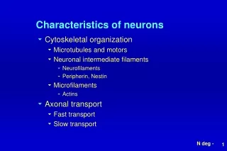
Dendrite (xs) Neurofilaments Microfilaments SER Microtubules Axon (ls) 40,000x MF MT NF 12,000x Neuronalcytoskeleton Microfilaments (4-6 nm) Neurofilaments (8-10 nm) Microtubules (20-25 nm) - Gives shape and support to the neuron - Facilitates intracellular transport - 25%-35% of total cell protein - Neuronal pathologies often involve the cytoskeleton either directly or indirectly
Sub-membrane distribution Dendritic spine Microfilaments Similar to microfilaments found in all cells, with neuron specific isoforms of the actin protein. Concentrated beneath the plasma membrane where they provide support for the plasma membrane and anchorage for membrane proteins. Prominent at nodes of Ranvier, post-synaptic densities. Form the core of dendritic spines. Node of Ranvier
-PO4 -PO4 -PO4 -PO4 -PO4 Neurofilaments Neurofilaments are intermediate filaments specific to neurons Provide support and stabilization; stable slow metabolic turnover. Phosphorylation gives additional stability in axons, especially in large, myelinated axons 50,000x
NF MT Longitudinal section Cross section Microtubules Similar to microtubules found in all cells, with neuron specific isoforms of tubulin proteins Provide the “intracellular highway” for transport
Intracellular transport Major synthetic site is the cell body Molecules and pre-formed organelles must be transported distally Return of materials for recycling Occurs in dendrites and axons, first studied in axons (axonal transport)
Fast transport (200-400 mm day) Anterograde = away from the cell body Neurotransmitter vesicles Vesicles for membrane insertion Mitochondria Retrograde = toward the cell body Recycled organelles Endocytic vesicles
Direction of transport depends on the orientation of the microtubules and different cargo motors.
Mechanism of fast transport Organelles (cargo) are transported along the “microtubule highway” by molecular motors which are ATPases: Kinesin - Anterograde transport, toward (+) end Dynein – Retrograde transport, toward (-) end Organelles bind to their specific motor move along the microtubule in a direction dictated by their motor.
Altered axonal transport in neurodegenerative diseases • Primary: Charcot-Marie-Tooth disease type 2 • Peripheral neuropathy with axonal degeneration. • Distal motor weakness, loss of touch sensation, wasting. • Develops in late childhood/early adulthood. Mutation in the kinesin isoform transporting mitochondria. • Secondary: Neurodegenerative disease (Huntington’s, ALS, Alzheimer’s and Parkinson’s); may result from abnormal intraneuronal inclusions. One of the most common inherited neurological disorders (37/100,000)
Presynaptic terminals
The chemical synapse Specialized adhesive contacts Site of neurotransmitter release by vesicle exocytosis (bouton) Transmitters diffuse across a gap which physically separates the cells. (synaptic cleft) Transmitters bind to receptors on the post-synaptic structure producing alterations in the state of membrane polarization (post-synaptic density). Structurally asymmetric, unidirectional transmission.
Pre-synaptic terminal (bouton) Neurotransmitter vesicles Mitochondria Actin (support and vesicle movement) Tubulo-vesicular membranes Active zone Mito Active zone
Transmitter vesicles Clear vesicles (30-50 nm) Dense core vesicles Clear vesicles – ACh, GABA, glutamate Dense core vesicles - catecholamines (epinephrine/norepinephrine) Large dense core vesicles – neuropeptides (substance P, somatostatin) Some terminals can contain more than one type Large dense core vesicles 70-200 nm
Pre-synaptic Synaptic cleft Post-synaptic Pre-synaptic 25 nm Active zone Active zone Synaptic cleft Postsynaptic density Postsynaptic density
Dendrite spines Small elevations (2 microns) on the surface of some dendrites. Receive the majority of excitatory input to the neuron. 20,000 spines/pyramidal neuron. 40% of the total surface area of some neurons. Inhibitory synapses occur more on proximal dendrites (shafts) and soma.
SER Spine apparatus Extension of the SER in the dendritic spine. Associated with actin filaments and ribosomes. Function is unclear, may be the site of synaptic protein synthesis and calcium homeostasis, both important in synaptic plasticity.
Normal Sensory Deprived Functional significance of dendritic spines Highly labile structures, form or collapse in seconds. Enriched environments increase the number of dendritic spines. Spine morphology and function are altered in mental retardation and Fragile-X syndrome.
Excitatory Inhibitory Excitatory vs inhibitory synapses Excitatory synapses (Gray type I) Spherical vesicles (e.g., acetylcholine, glutamate) Asymmetric – Conspicuous postsynaptic density Wide synaptic cleft Inhibitory synapses (Gray type II) Oval vesicles (e.g., GABA) Symmetric – Less conspicuous postsynaptic density Narrower synaptic cleft
Excitatory Inhibitory Excitatory Inhibitory
Classification of synapses based on postsynaptic structure Axo-dendritic and axo-spinous (most numerous) Axo-somatic Axo-axonic (on presynaptic terminals)
Synaptic vesicle synthesis and trafficking • 1. Delivery of synaptic vesicle components to plasma membrane • 2. Endocytosis of vesicle components to form new synaptic vesicles directly • (major pathway) • Endocytosis of synaptic vesicle components and deliver to tubulo-vesicular membranes • (minor pathway) • 4. Budding of synaptic vesicle • 5. Loading of neurotransmitter into synaptic vesicle • Secretion of neurotransmitter by exocytosis in response an action potential and calcium
Synaptic vesicle synthesis and trafficking Large dense core vesicles Vesicles and their contents are synthesized in the cell body Transported by fast axonal transport Synaptic and extra synaptic release (cell body and dendrites) Secreted by a regulated mechanism similar to peptide hormones; merocrine release Release in response to burst firing and increased intracellular calcium Release slower and more sustained than synaptic vesicles; no recycling
Chemical vs electrical synapses Chemical Slower, neurons are separating by a space Maximal plasticity Use chemical transmitters, sensitive to drugs and toxins Unidirectional (asymmetric) Most common Electrical Fast, provide ionic coupling via gap junctions Minimal plasticity Restricted distribution found in selected brain regions and throughout the retina
Halassa, et al., Trends Mol. Med., (2007) The Tri-partite synapse Astrocytes surround synapses in the CNS Scavenger function of ions and transmitter Each astrocyte contacts over 140,000 synapses Glutamate uptake and release