Download
1 / 26
260 likes | 366 Views
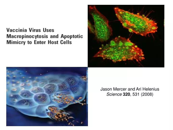
Jason Mercer and Ari Helenius Science 320 , 531 (2008). Infected child. Vaccinia virus and smallpox. L ive-virus vaccine against smallpox. Actual causative agent of smallpox – Variola virus 2 million deaths annually until 1959 WHO total vaccination programm Last fatal case – 1978

E N D
Jason Mercer and Ari Helenius Science 320, 531 (2008)
Infected child Vaccinia virus and smallpox Live-virus vaccine against smallpox Actual causative agent of smallpox – Variola virus 2 million deathsannuallyuntil 1959 WHO total vaccination programm Last fatal case – 1978 Completely eradicated by 1979 Immune responses generated from a Vaccinia virus infection protects the person against a lethal smallpox infection.
Vaccinia virus Poxvirus family dsDNA genome 190 kB over 200 proteins Vaccinia virus infection is very mild and is typically asymptomatic in healthy individuals Renewed interest in 21st century: -as a vector for immunization against other viruses. -due to concerns about smallpox being used as an agent for bioterrorism
eGFP E/L promoter eYFP A5 core protein Two infectious forms of vaccinia virus: Intracellular mature virus (MV) majority of infectious progeny, remains in the cell until cell lysis Extracellular enveloped virus (EV) surrounded by an additional, TGN-derived membrane Constructs used in this study EGFP-EXPRESS-MV EYFP-CORE-MV
MVs bind to filopodia and move towards the cell body Average speed: 1.05 ±0.38 µm/min
When MVs reach the cell body, large blebs are formed Infected cells Uninfected cells
Is there a correlation between formation of blebs and MOI? The quantity of blebbing cells but not the number of blebs per cell is MOI dependent
65% Without inhibitor 100μM Blebbistatin Is blebbing necessary for infection? Blebbistatin – an inhibitor of myosin II – dependent blebbing Infection Blebs formation Blebs formation is involved in productive viral entry
Which cellular factors are required for MV blebbing and entry? PAK1 (p21-activated kinase 1) Key regulator of the actin cytoskeleton, adhesion and cell motility Autoinhibited dimer Activated monomer PAK1 knockdown: siRNAs, dominant negative mutants, (AID) autoinhibitory domain expression
siRNA mediated knockdown of PAK1 68% 74% Do PAK1 siRNAs specifically block viral entry or downstream processes as well? Acid-mediated endocytosis by-pass assay, viral endocytosis is subverted, viral cores are delivered directly in cytosol under pH 5.0 PAK1 is necessary for prefusion events
Inhibition of infection by PAK1 autoinhibitorydomain (AID) 86 ± 4 % 8 ± 3 % 91 ± 6 % Number of infected cells eGFP-EXPRESS-MV infected cells – green mCherry-AID or mCherry-AID(L107F) transfected cells- red Actin -blue
PAK1 localized into membrane blebs during infection Cells, expressing eGFP-PAK1, actin is stained in red Uninfected 30 min post infection
Comparison of cellular factors involved in macropinocytosis, MV infectivity and MV inducedblebbing Perturbants and inhibitors pattern has shown that MV use macropinocytosis for entry
Professional phagocytes use macropinocytosis to engulf apoptotic bodies Phosphatidylserine (PS) - phospholipid component of the membrane In normal cells PS is kept on the inner-leaflet In apoptotic cells PS is exposed on the surface Nature Reviews Microbiology 2, 802-808, 2004
The MV membrane is enriched in phosphatidylserine MVs are similar with apoptotic bodies in size Blocking of cell surface or virion PS with annexin V
Inhibition of MV infectivity after treatment of virions with ANX5 Black bars – binding White bars - infectivity 90% PS is required for MV infectivity
Viral membrane delipidation by NP40 Without NP40 Green – eYFP-CORE-MV Red - actin 0.5% NP40 Delipidated virions can bindcells, but do not induce blebbing or entry
Virion lipid add-back Reconstitution of lipid bilayer on virions by incubation with PC based liposomes Liposome Phosphatidylcholine (PC)
Phosphatydlserine is exposed on the MV surface, can be extracted by NP40, and restoredwith PS liposomes. EYFP-A5-MV A647-Annexin V Merge Untreated Lipid extraction by 0.5% NP40 Lipid extraction by 0.5% NP40 and restoration with liposomes
The fate of vaccinia – infected cells: apoptosis or necrosis? Red – necrotic cells (PI) Green – apoptotic cells (PS-ANX5) MV spread is connected with apoptosis
Why do Vaccinia MVs use macropinocytosis andapoptotic mimicry to enter host cells? -Internalization of particlestoo big for other viral endocytic mechanisms. - Entry intothe different cell types, because PS-mediated clearance of apoptotic material is common to mostcells -Avoidance of the innate immune response