Download
1 / 17
180 likes | 342 Views
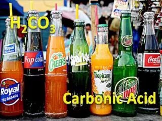
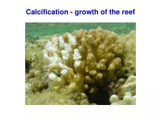
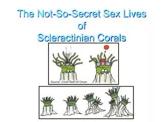
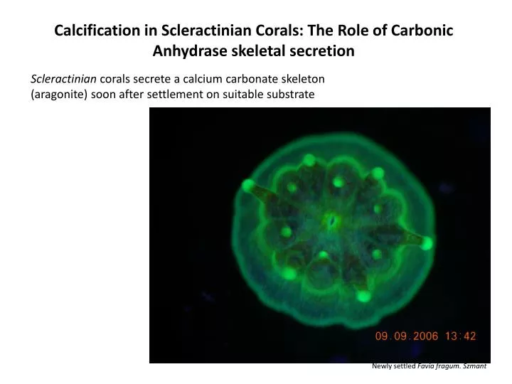
Calcification in Scleractinian Corals: The Role of Carbonic Anhydrase skeletal secretion. Scleractinian corals secrete a calcium carbonate skeleton (aragonite) soon after settlement on suitable substrate. Newly settled Favia fragum . Szmant. Brief Anatomy of a Scleractinian coral.

E N D
Calcification in Scleractinian Corals: The Role of Carbonic Anhydrase skeletal secretion Scleractinian corals secrete a calcium carbonate skeleton (aragonite) soon after settlement on suitable substrate Newly settled Faviafragum. Szmant
Brief Anatomy of a Scleractiniancoral Allemand et al. 2004
Calcification rates are increased during daylight and are reduced at night. Light enhanced calcification in the coral Stylophorapistillata. Is enhancement due to photosynthesis? Moya, A. et al. 2006
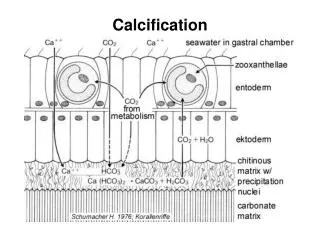
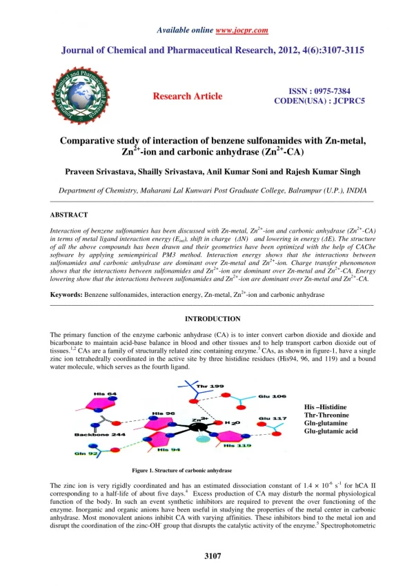
Proposed calcification mechanisms: Allemand et al, 2002 Calcium ion pathway in coral calcification has been well studied. Ca2+ ions are transported from the calicoblastic epithelial cells and into the subcalicoblastic area (site of calcification) via Ca2+ATPases.
Calcium ATPases localized to calicoblastic epithelium Zoccola et al, 2004
Goals of Study: • Confirm identity of new gene STPCA • Amplification of full mRNA sequence • Phylogenetic map • Characterize function of protein encoded by STPCA • Amino acid sequence alignment • Immunolocalization • Biochemical Characterization • Gene expression assay Carbonic Anhydrase in the Scleractinian Coral Stylophorapistillata AurelieMoyaet al. The Journalof Biological Chemistry. 2008 Sciencedaily.com Underlying question: How does CA fit into the mechanism of coral calcification?
Phylogenetic relationships of vertebrates and invertebratesα-CA sequences inferred from ML and Bayesian analyses STPCA groups with other Cnidarian CA sequences. Important because of symbiotic relationship and because of host of organisms that live within scleractinian skeletons Second AnthozoanCA grouping. Cytosolic form of Carbonic Anhydrase Groups with two other Anthozoan species. One is also a Scleractinian and the other is a Actinaria (non-calcifying, non-symbiotic, anemone) Moya A et al. J. Biol. Chem. 2008;283:25475-25484
Alignment of α-CA sequences from S. pistillata (STPCA), F. scutaria (FCAa), N. vectensis, Homo sapiens CAVI, XIV, I, II, A. elegantissima, and R. pachyptila • Signal Peptide present in STPCA sequence. • No GPI anchor in sequence • No evidence of trans-membrane regions (no hydrophobic regions in sequence). At the subcellular level, where are these enzymes found? Moya A et al. J. Biol. Chem. 2008;283:25475-25484
Immunolocalization of α-STPCA on the tissue slide of S. pistillata with carbonic anhydrase-specific antibody (anti-STPCA) or preimmune serum • A and B: DAPI, blue stain for nuclei. Anti-STPCA, orange dye • C: Control. DAPI and Preimmune serum STPCA shown to be localized to calicoblastic epithelium cells, adjacent to the skeleton. Little to no staining seen in other tissue types, especially the oral endoderm where much of the symbiotic algae resides. • SW: saltwater • OT: oral tissue • AT: aboral tissue • AE: aboral endoderm • CE: calicoblastic ectoderm • Co: coelenteron • Sk: skeleton Moya A et al. J. Biol. Chem. 2008;283:25475-25484
Western blot showing that A/STPCA protein is glycosylated Chimeric STPCA with polyhistidine tail attached to the C-terminus, and a normal STPCA gene treated with N-glycanase and run on a western blot. Results in both cases suggest glycosylation. Only anti-phosphoserine binds to enzyme in Western Blot. Results suggest serine and not threonine or tyrosine residues are site of phosphorylation. Amino Acid sequence analysis also shows predicted phosphorylation sites mostly on serine residues. Moya A et al. J. Biol. Chem. 2008;283:25475-25484
Kinetic parameters for carbon dioxide hydration reaction. STPCA activity is similar to secreted forms of human alpha carbonic anhydrase CA isoforms with similar kinetic parameters. All three are secreted forms of alpha carbonic anyhdrase Moya A et al. J. Biol. Chem. 2008;283:25475-25484 Mitochondrial isoforms Trans-membrane domain isoforms
STPCA gene expression significantly higher at night than during the day. • Corals kept in seawater system with a 12:12 hr light dark regime • Lighting provided by flourescent bulbs • Corals collected 12 hours apart, once during the day and once at night • Calcification rates are 2.6 times higher during the day than at night. • 2x upregulation of STPCA at night than during the day.
Summary of Findings: • Goals of Study: • Confirm identity of new gene STPCA • Amplification of full mRNA sequence • RACE amplification, BLAST confirm gene ID • Phylogenetic map • STPCA clustered with other cnidarian (secreted) CA isoforms • Characterize function of protein encoded by STPCA • Amino acid sequence alignment • No GPI anchor, sequence contains signal peptide, no hydrophobic regions found. • Immunolocalization • STPCA localized to areas involved with calcification (calicoblastic epithelium) • Biochemical Characterization • Enzyme is glycosylated and only serine residues are phosphorylated. • Gene expression assay • STPCA upregulated at night Underlying question: How does CA fit into the mechanisms of coral calcification? Photosynthesis?
Schematic model proposed for the function of CA in the calcification process Moya A et al. J. Biol. Chem. 2008;283:25475-25484 • Carbon dioxide is major carbon source for calcification. CO2 diffuses across CE membrane and into sub-calicoblastic epithelium where CA converts it into carbonic acid. • CA up-regulated at night to deal with decreased pH environment (low pH inhibits calcium carbonate reaction rate.
Critique: • STPCA role in light enhanced calcification. • Is night time up-regulation a biologically meaningful response to increased pH in the CE or indicative of a lag time between transcription and translation? Work done in Galaxeafascicularis, showing decreased night time pH levels in both the subcalicoblastic area and coelenteron. (Al-Horani et al, 2003)
Model fits well with calcification mechanism proposed by Allemand et al, 2004 DIC reservoir is carbon dioxide from CE cell mitochondria. Major difference is they proposed CA functioned in cytosol and carbonic acid transported to sub-calicoblastic region via a transport channel. Work done by Allemand et al, 2004 suggesting role of Carbonic Anhydrase in coral calcification. EZ is CA inhibitor. DIDS is a anion channel inhibitor.
Critique: • STPCA only functions in calcification and plays no role in photosynthesis. • Similar gene found in non-calcifying and non-symbiotic coral • Is lack of staining in other tissue layers a response to STPCA function of time of day? When were the corals used for staining collected? www.animalpicturesarchive.com www.genome.org