Download
1 / 24
270 likes | 1.06k Views
DIVERTICULOSIS. ADEL M. SALAMA, MD edited by Edward Warren, MD, Chair Geriatrics, Carolinas Campus, February 2012. Goals. Define diverticulosis/ itis . Discuss causes of diverticulosis/ itis. Discuss signs of diverticulosis/ itis . Describe the clinical evaluation of diverticulosis/ itis .

E N D
DIVERTICULOSIS ADEL M. SALAMA, MD edited by Edward Warren, MD, Chair Geriatrics, Carolinas Campus, February 2012
Goals • Define diverticulosis/itis. • Discuss causes of diverticulosis/itis. • Discuss signs of diverticulosis/itis. • Describe the clinical evaluation of diverticulosis/itis. • Discuss the treatment of diverticulosis/itis.
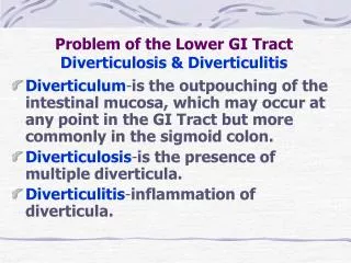
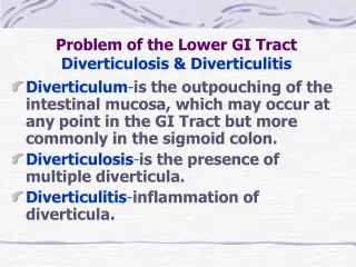
Definitions • Diverticuli are sacs: protrusions of the colonic mucosa. • Diverticulosis is the presence of diverticuli. • Diverticulitis refers to infection of diverticuli. • A typical colonic diverticulum is a pulsion diverticulum, where mucosa and submucosa herniate through the muscle layer covered by serosa. • Congenital diverticuli contain all layers. • Diverticular disease: diverticulosis & diverticulitis. • Symptomatic diverticular disease: bleeding, diverticulitis and complications of diverticulitis.
Epidemiology • Prevalence has increased 80 years ago it was 5-10% In 1969 it was 35-50% • Age dependent at age 40 <5-10% at age 60 30% at age 85 65%
Epidemiology (cont.) • Gender under age 50, more prevalent in MEN age 50-70, slight FEMALE preponderance above 70, marked FEMALE preponderance • Geographic variation In westernized nations, the prevalence is more left sided In Africa and Asia, the prevalence is more right sided *Japanese, after adopting a Western life style, have increase in prevalence of right sided diverticulosis
Etiology • Diet -- Total dietary fiber intake was noted to be inversely associated with symptomatic diverticular disease in a study involving 47,000 people • Physical activity -- The risk of developing symptomatic diverticular disease is inversely related to overall activity. • Obesity -- Overweight has been linked to the development of diverticulitis and diverticular bleed. • Colon cancer? -- One series of 7000 pts found excess number of colon and rectal CA in the first 2 years after the diagnosis of diverticular disease but not with prolonged follow up.
Pathophysiology • Point of weakness/ Vasa recta, Diverticula develop at these four well defined points around the circumference of the colon • Colon diameter- Law of Laplace P = k T /R Pressure is proportional to wall tension and inversely proportional to bowel radius. The sigmoid colon has the smallest diameter.
Pathophysiology (cont.) • Motility disorder -- Segmentation of the colon is a motility disorder in which segmental muscle contractions separate the lumen into chambers, this process is exaggerated in diverticulosis. The neural basis of this abnormality is unclear thought to be from upregulation of smooth muscle M3 receptors. • In geriatrics, there is declining colonic wall mechanical strength. • The acid solubility of collagen increases after age 40. • The cross linking of colonic collagen increases with age.
Pathophysiology (cont.) DIVERTICULITIS: The increased intraluminal pressure causes erosion, resulting in micro or macroscopic perforation. The inflammation is usually mild, walled off by pericolic fat. Obstruction or fistulae into adjacent organs with free perforation and peritonitis may occur. DIVERTICULAR BLEED: The vasa recta become draped over the dome, separated only from the lumen by mucosa. They become injured, leading to eccentric intimal thickening and thinning of the media with resultant wall weakness and bleeding. Right sided diverticulosis bleeds more due to wider domes.
Clinical manifestation • 70% Asymptomatic • 15-25% Develop diverticulitis • 5-15% develop diverticular bleed
Symptoms • Pain in 70%, for several days. Up to one half of pts have had previous, similar symptoms. • Nausea and vomiting 20-62% • Constipation 50% • Diarrhea 25-35% • Urinary symptoms 10-15% • Non specific complaint: cramping, bloating, irregular BM’s (mistaken as IBS). • Right sided diverticulitis can be misdiagnosed as acute appendicitis. • Diverticular bleeding is painless. Patients report passage of maroon or bright red blood (hematochezia). • It is rare for bleeding to coexist with diverticulitis.
Physical exam/Lab • Fever, low grade. • Tenderness, if generalized suggests perforation and peritonitis. • Mass • Leukocytosis/mild; if absent, no consequence since 45% had normal WBC. • LFT’s are normal. • Amylase can be mildly elevated in patients with perforation or peritonitis. • UA may reveal sterile pyuria.
Radiology CT with oral and IV contrast is the diagnostic test of choice. • Sensitivity 97%, Specificity100%, Positive predictive value 100%, Negative predictive value 98%. • Helps in diagnosis, assessment of severity, and planning therapeutic intervention. • Reports show • Increased soft tissue density within pericolonic fat 98%. • Bowel wall thickening 84%. • Soft tissue masses 35%. • Colonic diverticuli 84%.
Radiology (cont.) • Contrast enema • In the presence of perforation, (peritonitis) BARIUM enema is contraindicated. Use of water soluble contrast remains safe and demonstrate the site of the perforation. • Compression U.S • Better for patient with thin body. • Useful for rapid evaluation and diagnosis in ER setting. • Sensitivity 85-98% • Specificity 80-98% • Criteria: mural thickening > 4 mm in 5 cm segment.
Complications • Diverticulitis • Perforation • Abscess • Fistula • Obstruction
Treatment • Uncomplicated: 75% respond to medical management • Complicated: 25% require surgery
Uncomplicated • Outpatient: bowel rest (clear liquid for 2-3 days and antibiotics) • Inpatient: admission criteria include the eldery, immune suppression, multiple comorbidities, high fever and significant leukocytosis (NPO, IV fluids, and antibiotics) • Antibiotic: bacterial coverage includes gram negatives and anaerobes • ciprofloxacin 500 mg po bid and metronidazole 500 mg po tid for 7 - 14 days. • metronidazole for anaerobes (15 mg/kg IV and 7.5 mg/kg IV q6h) AND third generation cephalosporin for gram negatives like ceftriaxone 1 – 2 gm IV qd OR ciprofloxacin 400 mg IV q12h. • single agent can work: Ampicillin/sulbactam 3 gm IV q 6 hours OR piperacillin-tazabactam 3.375gm IV q 6h
Uncomplicated (cont.) • Dietary recommendations • Acute phase: clear liquids or NPO with IV fluids • Recovery phase: patients should be advised to consume a high fiber diet. • Long-term fiber supplementation may reduce the incidence of recurrence. • In a large cohort study, nut, corn and popcorn consumption did NOT increase risk of diverticulosis or diverticular disease.
Uncomplicated (cont.) • Monitor clinical course for outpatient treatment • Patient should be advised to call for increasing pain, fever, inability to tolerate fluids or fail to improve within 2-3 days…all are indications for hospitalization. • What next…treat aggressively in hospital…think of complication and treat accordingly…or think of other differential diagnosis like appendicitis, Crohn’s disease, ischemic colitis, ovarian cyst or torsion, ectopic pregnancy.
Uncomplicated (cont.) • 2 – 6 wks after resolution do a full evaluation of the colon to r/o neoplasia and to evaluate the extent of diverticulosis. • Prognosis after resolution • 30-40% remain asymptomatic • 30-40% will have cramps without diverticulitis • One third will have a second attack • Prognosis is worse with second attack • Complicated diverticulitis approaches 60% • Mortality is doubled • “Smoldering” diverticulitis occurs in some • The single strongest risk for missed cancer during colonoscopy was diverticular disease.
Complicated • Peritonitis Rx: Ampicillin 2gm IV q6h, Gentamicin 1 - 2 mg/kg IV q8h and metronidazole 500 mg IV q8h OR Piperacillin/tazobactam 3.375 mg IV q6h • Perforated diverticulitis has a mortality rate 6% for purulent peritonitis and 35% for fecal peritonitis • Obstruction: a primary concern is carcinoma • Abscess: percutaneous drainage permits elective single stage surgery in 60 - 80% of patients. The catheter is left in place until drainage is less than 10ml in 24 hours. This may take 30 days. • Fistula
Principles of surgery Surgery is advised after the first attack of complicated diverticulitis or after 2 episodes of uncomplicated. • Laparoscopic resection is best after acute diverticulitis has resolved and in patients with stage 1 or 2 disease according to the Hinchey’s classification. • stage 1 - pericolic or mesenteric abscess • stage 2 - walled-off pelvic abscess • stage 3 - generalized purulent peritonitis • stage 4 - generalized fecal peritonitis • Elective surgery can do resection and primary anastomosis in the same procedure. • Emergency surgery is typically a two-stage surgery: 1) resect diseased colon with a colostomy, 2) revise colostomy and reanastomose colon.
Small bowel diverticula • Duodenal small bowel diverticula occur in 2-5 percent of patients, most of them are extraluminal, near the papilla of Vater. They are usually asymptomatic and are associated with higher frequency of common bile duct pigment stones. • Jejunoileal diverticula occur in 1-2 percent of the general population. They are associated with disorders of intestinal motility and bacterial overgrowth, which can be treated with antibiotics.
References • Rafferty, shelliti practice parameters for sigmoid diverticulitis 2006 • Wong,WD, Wexner et al Dis colon and rectum 2000 • Stollman,NH, Raskindiagnosi and management of diverticular disease of the colon in adults 1999