Download
1 / 33
330 likes | 417 Views
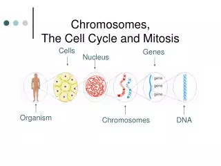
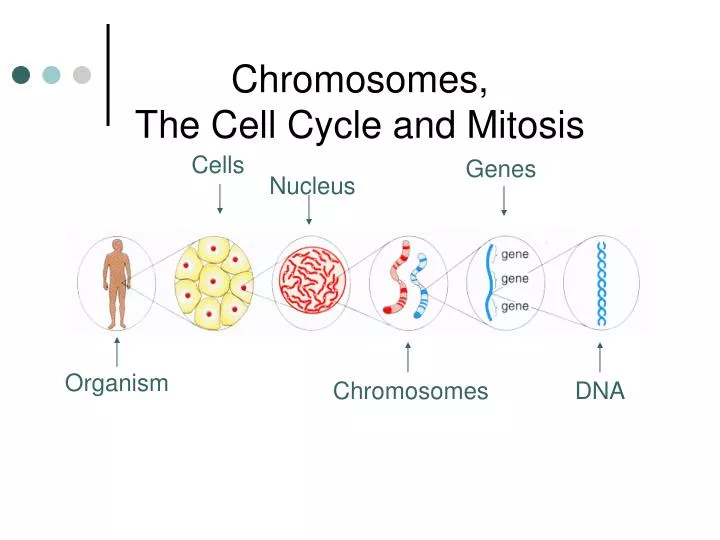
Chromosomes, The Cell Cycle and Mitosis. Cells. Genes. Nucleus. Organism. Chromosomes. DNA. Chromosome. A structure that forms when DNA wraps around proteins and coils up. Chromosomes only form right before the cell is going to divide. Each half of a chromosome is called a chromatid.

E N D
Chromosomes, The Cell Cycle and Mitosis Cells Genes Nucleus Organism Chromosomes DNA
Chromosome • A structure that forms when DNA wraps around proteins and coils up. • Chromosomes only form right before the cell is going to divide.
Each half of a chromosome is called a chromatid. Sister chromatids are identical to each other. Chromatids are joined at a centromere. Gene: Segment of DNA found on a chromosome. Each gene codes for a specific trait. Each chromosome has many genes on it. Chromosome Structure
Two Types of Chromosomes • Autosomes- chromosomes that do not help determine the gender of the individual (most of the chromosomes in your body) • Sex Chromosomes- in humans, they are X and Y; they determine the gender of the individual • XX = Female • XY = Male
Two types of Cells • Gametes- the reproductive cells (sperm for males, eggs for females). • Somatic Cells- all of the cells in the body that are not gametes. • Ex – skin cells, muscle cells, brain cells
Chromosome Number in Cells • Different cells can have different numbers of chromosomes depending on what type of cell they are. • Diploid VS Haploid • Diploid (2N)– two sets of chromosomes (one from mom, one from dad) • Somatic cells are diploid • Haploid (N or 1N)– one set of chromosomes • Gamete cells are haploid
Different organisms have different #’s of chromosomes. Ex: Drosophila melanogaster (fruit flies) : 8 Homo Sapiens (human): 46 Podocnemis uuiilis (turtle): 28
Homologous Chromosomes Karyotype - A karyotype is a picture of all of the chromosomes in the cell of an individual, taken right before the cell divides. Humans: 22 Pairs of Autosomes, 1 Pair of Sex Chromosomes
MUTATIONS Point Mutations (occur in the DNA on the genes) • Addition – adding on of extra nucleotides • A B C D A B B C D B. Deletion – subtraction of nucleotides • A B C D A C D C. Substitution – a different nucleotide is put in place • A B C D A B F D D. Translocation – nucleotides are switched around • A B C D C D A B
MUTATIONS Chromosomal Mutations (occur in the genes on the chromosome) • Some genetic disorders are characterized by having too many or too few chromosomes.
Trisomy 21 • Also known as “Down’s Syndrome” • Characterized as having an extra chromosome on the 21st pair. • Affects approximately 800 in 1,000 births • Maternal Age Risk at birth • 15 to 24 years 1 out of 1300 • 25 to 29 years 1 out of 1100 • 35 years 1 out of 350 • 40 years 1 out of 100 • 45 (and older) 1 out of 25
Klinefelter’s Syndrome • Characterized as a male with an extra “X” chromosome (XXY) • The condition exists in roughly 1 out of every 500 to 1,000 males • Extra “X” causes slight feminization (including small penis, tall physique, enlarged breast tissue and infertility)
Turner Syndrome • Characterized as a female with only one “X” chromosome (XO) • Turner syndrome occurs in about 1 out of 2,000 live births • Symptoms include webbed neck, drooping eyelids, short height and infertility
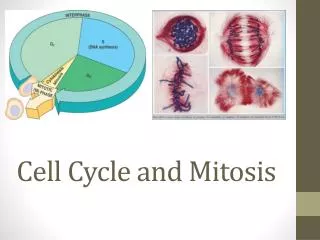
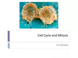
THE CELL CYCLE All somatic cells undergo the cell cycle in order to prepare for cell division (mitosis) • Mitosis is a form of asexual reproduction. The cells that form as a result of mitosis are clones (identical copies) of the original cell. • The reasons for mitosis are: • Growth of the organism • Repair of cells or replacement of “dead” cells
Stages of the Cell Cycle • G1 Phase • S Phase • G2 Phase • Mitosis • Cytokinesis
G1 (growth 1) Phase • First growth stage • Cell increases in size • Happens after cell division • Normal cell processes are occurring
Synthesis (S) Phase • Copying of all DNA • DNA is replicated • Chromosomes duplicated Chromosomes needs to be copied before a cell divides, so that each new cell has the correct amount of DNA and the correct number of chromosomes.
G2 (growth) Phase • The time between DNA synthesis (S) and mitosis (M). • Cell continues growing. • Proteins needed for cell division (mitosis) are produced.
Interphase – Resting Stage Interphase is the first 3 phases of the cell cycle together G1, S and G2 Cells carrying on normal activities • Chromosomes aren’t visible (not coiled up). • Normal cell metabolism and processes are occurring. • Occurs before mitosis. Includes phases of cell cycle “getting ready” for mitosis
Mitosis (M) and Cytokinesis Phases • Cell growth & protein production have stopped. • The cells energy is used to make 2 daughter cells (splitting of original cell into 2). • Mitosis – division of the nucleus into 2 nuclei in one cell. • Cytokinesis – division of cytoplasm, resulting in 2 new cells.
Cell Cycle Checkpoints The cell has checkpoints to ensure that each phase was completed correctly. G1: making sure the cell has grown enough and is able to divide. S/G2: making sure the DNA copied correctly and that all processes are ready for division. Mitosis (and Cytokinesis): making sure the cell divided correctly. Checkpoints not working and the cell dividing uncontrollably can lead to cancer.
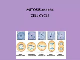
Stages of Mitosis • Prophase • Metaphase • Anaphase • Telophase
PROPHASE • DNA coils tightly & becomes visible as chromosomes. • Nuclearmembrane disappears • Centrioles migrate to poles • Centrioles: organelles that produce spindle • Spindle fibers begins to form
METAPHASE • Spindle fibers from centrioles attach to each chromosome. • Chromosomes line up in the middle of the cell.
ANAPHASE • Chromosomes are already separated into sister chromatids at the centromere. • Spindle fibers shorten and each chromatid is pulled to the opposite end of cell.
TELOPHASE • Separation of chromosomes into chromatids is completed • Nuclear membrane reforms. • Now have two nuclei in one cell • Chromosomes uncoil
Telophase – under the microscope Plant Animal
CYTOKINESIS • Cytoplasm division • Occurs after chromosomes separate and two nuclear envelopes reappear. • Forms two, identical daughter cells (they are identical to the original cell)
Check your chromosome numbers!!! Human Cell: Before G1 – 46 chromatids After S – 92 chromatids (46 chromosome pairs) After mitosis – 2 cells each with 46 chromatids!! Daughter cells are identical to the parent cell!!!