Download
1 / 56
950 likes | 2.8k Views
Clay Bundrick PGY-2 LSU Ophthalmology. Central Serous Chorioretinopathy. Presentation- Patient #1 . CC : sudden onset blurry vision OS HPI : 40 year BM c/o blurry central vision of L eye x 3 days symptoms vary from a black spot to central blurriness. + micropsia , no metamorphopsia

E N D
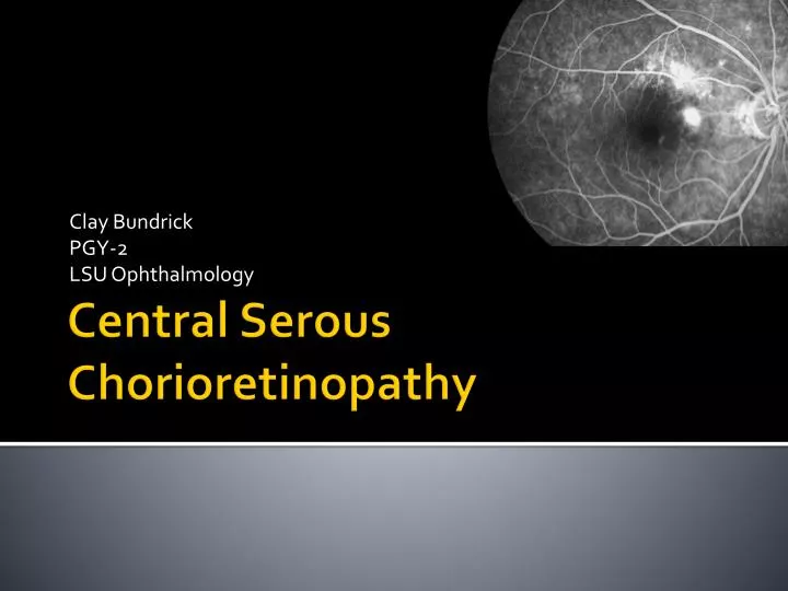
Clay Bundrick PGY-2 LSU Ophthalmology Central Serous Chorioretinopathy
Presentation- Patient #1 • CC: sudden onset blurry vision OS • HPI: 40 year BM c/o blurry central vision of L eye x 3 days • symptoms vary from a black spot to central blurriness. • +micropsia, no metamorphopsia • POH: none • FOH: none • PMH: HTN • Social Hx: carpenter, nonsmoker • Admits to elevated levels of stress secondary to death in family
Physical Exam, pt #1 • BP 133/86, HR 68/m, Temp 98.2F • Va- 20/20 od, 20/400 os • Ta- 14ou • Pupils- 31 reactive ou, no apd • Vf- grossly normal • SLE- wnl
Presentation- Patient #2 • CC: 49 yobm decreased vision osx 4 months • HPI: gradual onset blurry vision os with metamorphopsia and central blurring • POH: episode of blurry vision in right eye 4 years prior that resolved after 2-3 months • FOH: none
Past Medical History- Pt #2 • HTN- controlled on HCTZ • “slipped disk in back” • Rx with corticosteroid injections about every 3 months for several years • Last treatment with steroids 6 mo prior to pres • +s/o GERD/ gastritis, untreated undiagnosed etiology • No associated headaches
PE pt #2 • BP 127/80 HR 67 Temp 99F • scVA: 20/20 OD, 20/50 OS phni • BCVA: 20/25, MRx +2.25OS • Ta: 10 ou • SLE: wnl • Pupils reactive, no apd
CSCR • Idiopathic circumscribed serous RD • Usually confined to central macula • From fluid leaking through the RPE • NO signs of: • Inflammation • Accelerated HTN • Infiltration/ infarction of choroid/ RPE
Three types of CSCR • 1. Typical/ Classic CSCR • common • Younger pts • Acute localized RD • Mild to moderate loss of VA • Ass’d with one to a few focal leaks on FA
…. Three types • 2. Chronic CSCR • aka…Diffuse retinal pigment epitheliopathy • Widespread alteration of pigmentation of the RPE • From chronic shallow subretinal fluid • Likely to have h/o chronic steroid usage • 3. Bullous RD secondary to CSC • rare
Demographics • Historically thought to be found most commonly in men aged 20-45 years old • More recent studies have found… • mean age 45-51 years old • Males: females 3:1 (prev thought to be 6:1) • Whites, hispanics, asians >> blacks • Refuted by some • More severe in hispanics and asians with extensive bullous changes
Associations • The big players • Corticosteroids • Stress • Type A personality
Associations • Increased cortisol levels • Cushing’s, inhaled nasal, epidural • Organ transplants rx with immunosuppressants • Pregnancy • Steroids reduce nitric oxide levels (vasodilators) • +htn, +increased catecholamines • Hypothesize that adrenergic effectors cause vasoconstriction and alter choroidalhemodynamics
Associations • Helicobacter pylori: • significantly higher prevalence of H pylori infection in CSCR patients compared to age matched populations • Link: possible immune mechanism, based on a "molecular mimicry" between pathogenic antigens expressed on the bacterium and homologous host proteins
Associations Methlenedioxymethamfetamine (ecstasy)
pathophysiology • Well circumscribed serous detachment of sensory retina with RPE changes +/- RPE detachment • Mechanisms: • - Altered RPE barrier and pumping functions • - Choroidal microcirculation abnormalities: • -localized vasoconstriction, hypermeability • -impaired fibrinolysis • ?occlusion of choriocapillaries with decreased fovealchoroidalblood flow
Biomicroscopic findings • Solitary round shallow RD in PP • Avg size is 2 DD’s • Serous PEDs common- • more orange elevation with darker rim • RPE cells and BM’s detach from Bruch’s **PED’s with turbid subretinal fluid or blood most likely from CNV --either OCCULT CNV or polypoidalchoroidal CNV
OCT • Patient # 1 Patient #2
FA- 2 patterns of leakage • Smokestack (~only 10%), most dramatic • First arises superiorly • Then plumes out laterally • Usually associated with larger areas of RD
FA- classic CSCR- 90% • One or more hyperfluorescent leaks at the level of the RPE • Early- focal dot like hyperfluorescence this is leakage of dye from the choroid through the RPE
FA- classic CSCR • Later • Increasing size and intensity of the hyperfluorescent dot
Where is the focal leak? • Focal leaks more common nasally and superiorly • Most leaking points are in a ring 1mm wide starting 0.5mm from the center of the fovea • Can occur further than 3mm from the FAZ (11.8%) • IN the fovea <10% of cases • Within papillomacular bundle 20-25% cases
ICG Findings of Classis CSCR • Multifocal areas of choroidal vascular hyperpermeability • Best seen in mid phases • These hyperpermeability regions may persist in chronic csceven when there is no active leakage on FA • Important tool to differentiate from occult CNV
Chronic CSC • Diffuse Retinal Pigment Epitheliopathy • Widespread alteration of pigmentation of decompensating RPE in the posterior pole • Related to chronic presence of subretinal fluid • Usually worse- VA loss more pronounced
Chronic CSCR • - widespread disease • Retinal thinning/ cystic changes • 3 RPE changes • 1. atrophy, thinning • 2. Focal hyperpigmentation • 3. Hyperplasia c bone spicules • Subretinal lipid can be seen • Masquerades as occult CNV • Different rx, important to differentiate
FA chronic CSCR- patient #2 As CSCR becomes chronic it loses itspinpoint character and becomes diffuse
Bullous RD 2° to CSCR • Rare • Numerous exuberant leaks • Multiple PED’s • Bullous rd extending into inferior periphery of hundus • 84% bilateral • 36% as progression from classic I
Differential Diagnosis • CNV • Tumors • Inflammatory conditions • Vascular pathology • Rhegmatogenous RD
Differential…. CNV • Most important to differentiate • Particularly occult CNV • BOTH • Neurosensensory detachments, PEDs • Hyperpigmentation, RPE atrophy • Subretinal deposits of fibrin and lipid • CNV • Thickened RPE with notched PEDs • Subretinal, subpigment epithelial blood
DDx…. CNV • Thickened RPE with notched PEDs • Subretinal, subpigment epithelial blood
Angiography to differentiate CNV -Unilateral, unifocal area of hyperfluorescencethat usually shows progressively increasing contrast with surrounding choroid in the later phases May be helpful to repeat fa 2-3 weeks later as cnv may become more apparent with time
DDx… Ocular Histoplasmosis • OHS- punched out chorioretinal scars
DDx continued • Tumors • Leukemia • Amelanotic melanoma • Metastasis • Infiltrative lesions different color than surrounding choroid • Thickening on ultrasound • No serous PED
DDx continued • Inflammatory conditions with serous RD’s • Posterior Scleritis • Harada’s disease • Other signs of inflammation- vitritis and iritis • Stained optic nerve head on FA • Thickened choroid
Fluorescein angiography of the left eye in a patient with Vogt-Koyanagi-Harada disease. Mid phase is shown on the left with multiple areas of hyperfluorescence at the level of the retinal pigment epithelium. Late phase of the same angiogram (right) reveals multiple placoid areas of hyperfluorescence at the level of the retinal pigment epithelium and pooling of dye in the areas of serous detachment.
DDx continued • Optic Nerve Pits • May have serous detachment of macula • Generally a bilateral detachment caused by retinoschisis in the macula • No leaks at level of RPE
DDx continued • Rhegmatogenous RD • Elevation of macula can mimic cscr • Associated retinal hole or tear • No leaks on FA
DDx Continued • Vascular Disoders • Malignant HTN • Differentiated by presence of systemic HTN • Elschnig’s spots • Shifting fluid • Retinal vasc changes
Natural Course • Most spontaneosly and completely resolve • Average resolution time is 3 months • 94% eyes had 20/30 Va after 23 months • Many still have complaints however: • Decreased color vision • Relative scotomas • Micropsia, metamorphopsia, nyctalopia • Even though the neurosensory detachment resolves there has still been damage to photoreceptors, irregular rpe pigmentation, atrophy and subretinalfibrosis
Natural Course • 40-50% risk of recurrence and being chronic • Risk CNV from previous CSCR is considered small (< 5%) but has an increasing frequency in older patients diagnosed with CSCR • Ultimately 5-10% develop DRPE • STUDY: • reviewed a subset of patients who presented with a severe variant of CSCR over a mean follow-up period of 10.6 years. • These patients were characterized by multifocal lesions and bullousRDs with shifting fluid and fibrin deposition. • During the follow-up period • 52% of patients experienced recurrences of CSCR ranging from 1-5 episodes. • However, 80.4% of eyes (n=46) returned to a visual acuity of better than 20/40 • and 52% returned to a visual acuity of 20/20 or better. • Eventually, patients reached a state of quiescent disease
Clinical Prognosticators • - Prospective case series of 46 eyes, mean f/u 22.8 months • - Statistically significant correlation between baseline BCVA , duration of symptoms and final BCVA • - Shorter periods of subfoveal fluid correlated with better VA. • - Both baseline and final BCVA were strongly correlated with foveal thickness after fluid resolution • - Angiographic pattern were not significantly correlated with better VA.
Treatment • No medical therapy shown to be effective…. Yet? • Beta blockers, barbiturates, ketoconazole not shown to be effective • Laser Photocoagulation • Photodynamic Therapy
Laser • Goal to reduce leakage through RPE • Apply laser to site of leakage seen on FA • Shorten duration of macular detachment • Does not seem to affect final VA • DRPE • Thermal grid laser to small leaks • Decreases subretinal fluid • No longterm benefit in va • Inconclusive evidence on its effect on recurrence rates
Laser…. • Bullous variant- no advantage • Side effects of photocoagulation include • CNV • Scotoma • RPE scar expansion
When to laser? • Symptoms >4 months • Leakage sites located >375um from fixation • History ofcscr in fellow eye • Need or desire for treatment • If leak is located well away from central macula no need to be as hesitant
Methods of photocoagulatoin • To the leakage site with low-intensity • Spot size 100-200microns • Power of 100-150mW • Application time of 0.1-0.2s • Laser uptake depends on • Amt of fluid • Degree of pigmentation/ detachment of RPE • Important to obtain only a dull gray coagulation to avoid secondary CNV
Photodynamic Therapy • Photosensitizers accumulate in and are retained by proliferating tissues • When stimulated by appropriate λ… • active O2/ free radicals are generated • Resulting in photochemical damage to these cells • Approved for subfoveal CNV • Extensively studied for neoplasiarx
Photodynamic Therapy • May be useful for DRPE • Challenging to treat bc of widespread indistinct leaks • May be useful in acute csc but cost of verteporpfin is limiting • Generally causes subretinal fluid to decrease or resolve completely