Download
1 / 25
340 likes | 893 Views
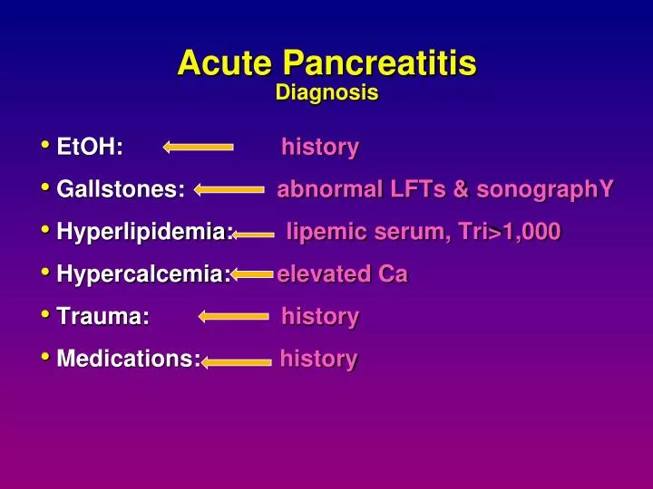
Acute Pancreatitis Diagnosis. EtOH : history Gallstones: abnormal LFTs & sonographY Hyperlipidemia : lipemic serum, Tri>1,000 Hypercalcemia : elevated Ca Trauma: history Medications: history.

E N D
Acute PancreatitisDiagnosis • EtOH: history • Gallstones: abnormal LFTs & sonographY • Hyperlipidemia: lipemic serum, Tri>1,000 • Hypercalcemia: elevated Ca • Trauma: history • Medications: history
Abdominal Exam • Abdominal tenderness and rigidity • Bowel sounds decreased • Palpable upper abdominal mass Acute fluid collections and pseudocysts • Skin Exam • Erythematous skin Nodule (Subcutaneous Fat Necrosis) • Cullen's Sign (periumbilical discoloration) • Turner's Sign (flank discoloration) * due to exudation of blood-stained fluid into the subcutaneous tissue, usually 72 h into the illness.
Acute PancreatitisClinical Manifestations PANCREATIC PERIPANCREATIC Adjacent viscera: SYSTEMIC Mild: edema, inflammation, fat necrosis Severe:phlegmon, necrosis, hemorrhage, infection, abscess, fluid collections Retroperitoneum, perirenal spaces, mesocolon, omentum, and mediastinum ileus, obstruction, perforation Cardiovascular: hypotension Pulmonary: pleural effusions, ARDS Renal: acute tubular necrosis Hematologic: disseminated intravascular coag. Metabolic:hypocalcemia, hyperglycemia
Serum Amylase elevated Nonspecific Returns to normal in 48-72 hours Normal amylase does not exclude pancreatitis Level of elevation does not predict disease severity Serum Lipase elevated Specific for pancreatic disease Returns to normal in 7-14 days Diagnosis: Biochemical
White Blood Cells increased to 15k-20k Hypertriglyceridemia (15%) liver Function Tests (ALP) (AST) ,elevated (LDH) elevated (Poor prognosis) Hyperglycemia Albumine (Poor prognosis) Serum Electrolytes Hypocalcemia (25%) Diagnosis: Biochemical
Another criteria often used to assess the severity of pancreatitis is the (APACHE-II) . Acute Physiology And Chronic Health Evaluation age and vital signs Specific laboratory parameters, Chronic health status The main advantage is the immediate assessment of the severity of pancreatitis. A score of eight or more at admission is usually considered indicative of severe disease
Predictors of Severity • Why are they needed? • Appropriate triage & therapy • compare results of studies of the impact of therapy • When are they needed? • optimally, within the first 24 hours • Which is the best?
AT ADMISSION Age > 55 years WBC > 16,000 Glucose > 200 AST > 250 IU/L LDH > 350 IU/L WITHIN 48 HOURS HCT drop > 10% BUN > 5 Arterial PO2 < 60 mm Hg Base deficit > 4 mEq/L Serum Ca < 8 Fluid sequestration > 6L Ranson CriteriaAlcoholic Pancreatitis Number <2 1% 3-4 16% 5-6 40% 7-8 100% Mortality
Glasgow CriteriaNon-alcoholic Pancreatitis • WBC > 15,000 • Glucose > 180 • BUN > 16 • Arterial PO2 < 60 mm Hg • Ca < 8 • Albumin < 3.2 • LDH > 600 U/L • AST or ALT > 200 U/L
CT Severity Index Balthazar et al. Radiology 1990.
Useful markers of severe disease. • Pleural effusion • BMI (High body mass index) • Necrosison contrast-enhanced CT-SCAN • CRPlevel greater than 150 mg/L at 48 h • Infection of the necrotic tissue after the first week of illness is the major determinant of later outcome.
CT-guided percutaneous fine-needle aspiration of the pancreatic tail
Pancreatic Cancer Pancreatic cancer is one of the most lethal malignancies. An estimated 32,300 patients will die of pancreatic cancer in year 2006. The tenth most common malignancy in the United State. Despite recent advances,inpathology, molecular basis and treatment, the overall survival rate remains 4% for all stages and races. Palliative care represents an important aspect of care in patient with pancreatic malignancy. Identifying and treating disease related symptomology are priorities. Common problems include pain, intestinal obstruction, biliary obstruction, pancreatic insufficiency, anorexia-cachexia and depression.