Download
1 / 19
240 likes | 632 Views
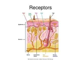
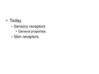
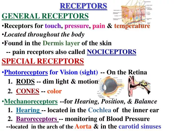
RECEPTORS. Found in the Dermis layer of the skin. Photoreceptors for Vision (sight). --for Hearing, Position, & Balance. GENERAL RECEPTORS. Receptors for touch , pressure , pain & temperature. Located throughout the body. -- pain receptors also called NOCICEPTORS.

E N D
RECEPTORS • Found in the Dermis layer of the skin • Photoreceptors for Vision (sight) --for Hearing, Position, & Balance GENERAL RECEPTORS • Receptors for touch, pressure, pain & temperature • Located throughout the body -- pain receptors also called NOCICEPTORS SPECIAL RECEPTORS -- On the Retina 1. RODS -- dim light & motion 2. CONES -- color • Mechanoreceptors 1. Hearing -- located in the Cochlea of the inner ear 2. Baroreceptors-- monitoring of Blood Pressure --located in the arch of theAorta & in the carotid sinuses
3. Proprioceptors --Located in muscles, tendons, joints & the semicircular canals of the inner ear --Provide information on position, balance & movement of limbs 1. Located in the digestive tract & in the hypothalamus 2. Monitor for changes in the concentration of body fluids (Electrolytes) • Chemoreceptors -- for Taste, Smell, O2 & CO2 levels 1. Taste --taste buds located on the tongue fissures 2. Smell --olfactory cells located in roof of the nasal cavity 3. Receptors for O2 & CO2 located in arch of the Aorta & carotid sinuses • Osmoreceptors --4 types (Bitter, Sour, Salty, & Sweet)
DEFINITIONS -- Receptors adjust themselves so that they no longer respond to the stimulus as they did in the beginning. -- Some neuron connect impulses from the skin & the viscera to send to the brain -- The brain cannot differentiate, so assign pain to the skin Gallbladder pain may be felt in in the shoulder Kidney pain may be felt in the lumbar (flank)region ADAPTATION REFERRED PAIN
Clear, transparent, anterior coat of the eye Part that is replaced in a corneal transplant Watery fluid & helps Lens Cornea maintain curve of cornea & carries nutrients Aqueous to the cornea & lens humor thickness is adjusted to focus light -- lens thins or flattens out Suspensory for distance ligament -- lens thickens or rounds up for near vision Ciliary body EYE -- hold Lens in place & help it to change shape
-- the change of direction of a light ray as it passes from one medium to another -- coordinated eye changes to enable us to focus on on objects Refraction (bending) Accommodation
Jelly-like substance that give the & aids in refraction eye shape If lost cannot be replaced causing blindness as retina falls forward Cornea Removal of the eyeball is calledEnucleation absorbs stray light Retina rays & nourishes Choroid the retina Sclera opaque back portion of Vitreous humor the Cornea Rods -- functions in dim light & senses motion Cones -- functions in bright light & is sensitive to 3 colors Red Green Light blue
Fovea centralis Retinal artery & vein Optic Optic nerve -- Cranial Nerve II carries visual impulses from the rods disc & cones to the Occipital Lobe Papilledema (Blind Spot) -- area in which there is no rods or cones --swelling & inflammation of the optic nerve Area where the vascular bed can be directly examined -- area that contains a large amount of cones, so it is the point of most acute vision
Muscles of the Eye Lens Iris -- regulates amount of light entering the eye Pupil -- distortion of the curvature of the cornea Intrinsic muscles-- inside the eye Colored portion of the eye -- rounds up or flattens to view objects Myopia -- nearsightedness Hyperopia -- far-sightedness Astigmatism Presbyopia -- old-sightedness
Extrinsic Muscles TrochlearAbducens & 6 muscles attach to the skull bones & the sclera to allow for coordinated movement Nerves for the Extrinsic Muscles -- supplies one extrinsic muscle each Oculomotor -- supplies 4extrinsic muscles
Protection of the Eye Conjunctiva Lines the eyelid & cornea Destroys pathogens Lacrimal Tears film across the eye ducts in the direction of the nose Lacrimal gland -- Skull bones -- Eyelid & eyelashes --eyelashes sense particles -- sebaceous glands help lubricate the eye (Sty -- inflammation of one of these glands) Produces tears Keeps the eyeball moist
EAR Pinna Made of cartilage Helps collect sound waves Not necessary for hearing External Auditory Canal Tympanic Membrane External Structures Also called the Auricle which captures foreign material & protects the ear Sound vibrations are first picked up here In small children, it is chiefly made of cartilage
Middle Ear It’s a cavity filled with air & containing Ossicles Incus Stapes Equalizes pressure Malleus Lies between the tympanic membrane & Incus Shaped like a hammer Eustachian Tube Connects middle ear to the throat Easy route for infection Shaped like an anvil & lies between the Malleus & Stapes Attaches to the Oval window, which is the dividing line between Middle & Inner ear These bones are joined in such a way that sound waves are amplified 20x by the time they reach the oval window
Inner Ear -- vibrations enter here through the Oval window Vestibule Contains Organ of Corti, the site for the sensory receptor cells (Hair Cells) Cochlea Impulses are picked up here and sent to the 8th Cranial nerve Vestibulocochlear Semicircular canal Vestibulocochlear nerve Osseous Labyrinth Functions in hearing Base of the canal contains 2 sacs with hair cells that deal with head position & equilibrium
Membranous Labyrinth membrane bone -- is the fluid inside the Membranous Labyrinth perilymph endolymph Within the Bony Labyrinth is a Membranous Labyrinth The inner ear containfluid Between the Bony labyrinth and the Membranous labyrinth is a fluid called: Perilymph Endolymph
Balance & Equilibrium Senses eye movement & head position Also send impulses to Vestibulocochlear nerve & then to the Cerebellum
Hair Cells Basilar Membrane Tectorial Membrane Organ of Corti Endolymph
Sound Waves Vibration of the Tympanic Membrane Vibration of Middle Ear bones Vibration of Oval Window Fluid Movement Vibration of within Cochlea Round Window Vibration of the Dissipation Basilar Membrane of Energy Electrical Stimulus Impulse sent to Bending of Hairs of picked up by the the Temporal Lobe Receptor Cells of Organ of Vestibulocochlear Corti starts of the Cerebral the Electrical Nerve Cortex Stimuli
Otitis Media -- inflammation or infection of the middle ear Otosclerosis -- immobility of an ossicle usually the stapes Treatment is a Stapendectomy HEARING LOSS Conductive Deficit --Causes: Cerumen (ear wax) Foreign bodies Perforation of the tympanic membrane Treat underlying cause
Cilia (hair cells) on the receptors have worn away is the most frequent cause of nerve deafness Nerve Deficit -- Damage to the 8th Cranial Nerve --Causes: Prolonged exposure to loud noises Drugs like Streptomycin; Neomycin Cochlear atrophy German measles in the first trimester Mumps