Download
1 / 40
500 likes | 932 Views
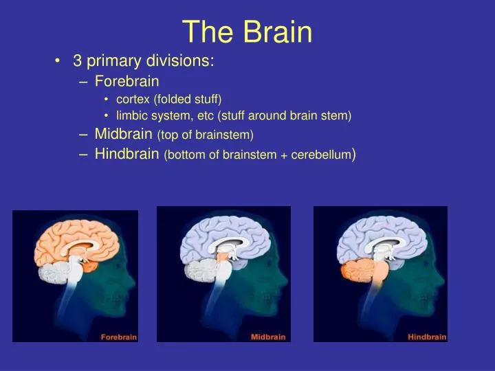
The Brain. 3 primary divisions: Forebrain cortex (folded stuff) limbic system, etc (stuff around brain stem) Midbrain (top of brainstem) Hindbrain (bottom of brainstem + cerebellum ). Hindbrain. Medulla Pons Cerebellum. Pons. Medulla. Cerebellum.

E N D
The Brain • 3 primary divisions: • Forebrain • cortex (folded stuff) • limbic system, etc (stuff around brain stem) • Midbrain (top of brainstem) • Hindbrain (bottom of brainstem + cerebellum)
Hindbrain Medulla Pons Cerebellum Pons Medulla Cerebellum http://www-unix.oit.umass.edu/~psyc335c/lectures/hindbrain.gif
Medulla: Controls vital reflexes: breathing, heart rate, vomiting, salivation, coughing, sneezing - Via cranial nerves Damage to medulla can be fatal Large doses of opiates can be fatal b/c suppress activity of medulla…why…?...b/c receptors there!
Pons: Also has cranial nerves Location of axon decussation (where axons cross from one side of the brain to the other…so left brain controls right body and vice versa) Reticular formation: motor control, arousal, consciousness
Midbrain: Cerebral aqueduct More cranial nerves Superior colliculus (visual info) Inferior colliculus (auditory info) Substantia nigra: dopamine-producing cells, structure that is lost in Parkinson’s Disease http://en.wikipedia.org/wiki/Midbrain
Brainstem Medulla Pons Midbrain Some forebrain structures
Senses: Information comes in the cranial nerves and eventually ends up in the cortex
Cranial Nerves Table 4.4, page 87 Olfactory nerve: Smell http://www.besthealth.com/besthealth/bodyguide/reftext/images/cranial_nerves.jpg
Cranial Nerves Table 4.4, page 87 Optic nerve: Vision http://www.besthealth.com/besthealth/bodyguide/reftext/images/cranial_nerves.jpg
Cranial Nerves Table 4.4, page 87 Occulomotor nerve: Eye movement, pupil constriction http://www.besthealth.com/besthealth/bodyguide/reftext/images/cranial_nerves.jpg
Cranial Nerves Table 4.4, page 87 Trochlear nerve: Eye movement http://www.besthealth.com/besthealth/bodyguide/reftext/images/cranial_nerves.jpg
Cranial Nerves Table 4.4, page 87 Trigeminal nerve: Skin senses from face Jaw muscles for chewing and swallowing (muscles of mastication) http://www.besthealth.com/besthealth/bodyguide/reftext/images/cranial_nerves.jpg
Cranial Nerves Table 4.4, page 87 Abducens nerve: Eye movements http://www.besthealth.com/besthealth/bodyguide/reftext/images/cranial_nerves.jpg
Cranial Nerves Table 4.4, page 87 Facial nerve: Taste Facial expressions Crying Salivation Dilation of head’s blood vessels http://www.besthealth.com/besthealth/bodyguide/reftext/images/cranial_nerves.jpg
Cranial Nerves Table 4.4, page 87 Acoustic nerve: Aka vestibulocochlear or statoacoustic Hearing Equilibrium http://www.besthealth.com/besthealth/bodyguide/reftext/images/cranial_nerves.jpg
Cranial Nerves Table 4.4, page 87 Glossopharyngeal nerve: Taste Swallowing Salivation Throat movements during speech http://www.besthealth.com/besthealth/bodyguide/reftext/images/cranial_nerves.jpg
Cranial Nerves Table 4.4, page 87 Vagus nerve: Sensation from neck and thorax Control of throat, esophagus, larynx Parasympathetic nerves to stomach, intestines, etc http://www.besthealth.com/besthealth/bodyguide/reftext/images/cranial_nerves.jpg
Cranial Nerves Table 4.4, page 87 Spinal accessory nerve: Aka Accessory nerve Neck and shoulder movements http://www.besthealth.com/besthealth/bodyguide/reftext/images/cranial_nerves.jpg
Cranial Nerves Table 4.4, page 87 Hypoglossal nerve: Muscles of tongue http://www.besthealth.com/besthealth/bodyguide/reftext/images/cranial_nerves.jpg
Cranial nerve signs help determine the location of a lesion in the brain • Essential element in clinical neuroanatomy • Neurological exam: http://www.vhct.org/case1799/neurologic_examination.shtml • Example: patient is asked to stick out tongue. If the tongue deviates to the left, the lesion involves the nucleus of the left hypoglossal nerve.
Nerve key Nerve Type of function On Optic Some = sensory Old Olfactory Say Olympus Occulomotor Marry = motor Towering Trochlear Money Tops Trigeminal But = both (S&M) A Abducens My Fin Facial Brother And Acoustic* Says German Glossopharyngeal Bad Viewed Vagus Boys Some Spinal accessory** Marry Hops Hypoglossal Money * Acoustic-vestibulocochlear, stateocochlear ** Spinal accessory = accessory
Forebrain • Thalamus • Hypothalamus • Pituitary gland • Basal ganglia • Basal forebrain • Hippocampus • Limbic system
Thalamus: Relay station for all sensory info on its way to brain (except olfactory info) Many specialized nuclei (ex: LGN, MGN…don’t have to know these!)
Hypothalamus Communicates with pituitary gland to alter hormone release Involved in feeding, drinking, temperature regulation, sexual behavior, fighting, arousal (activity level)…4 Fs
Pituitary gland Endocrine gland (hormone producing) Attached to base of hypothalamus by stalk Makes and releases hormones into bloodstream
Basal Ganglia Motor control, but also memory and emotional expression Lose dopamine neurons in SN Parkinson’s Disease Lose dopamine neurons in caudate & putamen Huntington’s chorea http://www.uni.edu/walsh/basalganglia-2.jpg Don’t memorize image!!! Just understand that this is a very complex system! thalamus.wustl.edu/ course/cbell6.gif
Basal forebrain Anterior and dorsal to hypothalamus Important for arousal, wakefulness, attention Lose cells in nucleus basalis decreased attention & intellect (AD, PD) http://memorylossonline.com/summer2003/glossary/basalforebrain.jpg
Hippocampus Memory formation HM: temporal lobes removed for intractable epilepsy no longer formed new memories http://www.hermes-press.com/Perennial_Tradition/hippocampus.gif http://www.umassmed.edu/bnri/graphics/crusiofig1.gif
Limbic System important for motivated & emotional behaviors (eating, drinking, sexual activity, aggressive behavior)
Ventricles Contain cerebrospinal fluid (CSF) CSF reabsorbed into blood vessels, so continuous turnover Protective Reservoir for hormones, nutrients http://mywebpages.comcast.net/epollak/PSY255_pix/ventricles.PNG
Ventricle size can indicate problems • Enlarged ventricles as in Alzheimer’s patients (cell loss). • Lack of ventricles due to tumors etc.
Cortex • 2 hemispheres • Communicate via corpus callosum & anterior commisure • 4 lobes http://pegasus.cc.ucf.edu/~Brainmd1/brmodelc.gif http://www.urmc.rochester.edu/neuroslides/slides/slide201.jpg http://trc.ucdavis.edu/mjguinan/apc100/modules/Nervous/grosscns/images/brain10.jpg
The lobes of the cortex • Frontal • Thinking • Prefrontal cortex • Planning • Working memory • Socially appropriate behavior • Delayed-response task • Lobotomies • Primary motor cortex • Broca’s aphasia
The lobes of the cortex • Parietal • Sensing • Primary sensory cortex
The lobes of the cortex • Temporal • Spoken language comprehension • Wernike’s aphasia • Hearing • Vision • Movement perception • Face recognition • Emotional motivational behavior
The lobes of the cortex • Occipital • Vision • Primary visual cortex • Damage causes “cortical blindness”
Evolution of Gene Related to Brain's Growth • A gene that helps determine the size of the human brain has been under intense Darwinian pressure in the last few million years. • It has changed its structure 15 times since humans and chimps separated from their common ancestor. • Evolution has been particularly intense in the five million years since humans split from chimpanzees Changes in the architecture of the ASPM protein over the last 18 million years are correlated with a steady increase in the size of the cerebral cortex (2002) Dr. Bruce T. Lahn at U. Chicago. A disrupted form of this gene was identified as the cause of microcephaly (people born with an abnormally small cerebral cortex).
Functions • Forebrain • the cool stuff (thinking, perceiving, big part of emotion) • Midbrain • sensory pathways • Hindbrain • motor control, reflexes (breathing, heart rate, etc)