Download
1 / 14
260 likes | 597 Views
Medical Imaging Modalities. Methods In Medical Image Analysis— Spring 2013 BioE 2630 (Pitt) : 16-725 (CMU RI) 18-791 (CMU ECE) : 42-735 (CMU BME) Dr. John Galeotti. Anatomical Axes. Superior = head Inferior = feet Anterior = front Posterior = back Proximal = central

E N D
Medical Imaging Modalities Methods In Medical Image Analysis—Spring 2013 BioE2630 (Pitt) : 16-725 (CMU RI) 18-791 (CMU ECE) : 42-735 (CMU BME) Dr. John Galeotti
Anatomical Axes • Superior = head • Inferior = feet • Anterior = front • Posterior = back • Proximal = central • Distal = peripheral
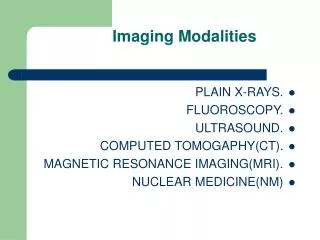
Imaging Modalities • Camera: Microscope, Endoscope, etc. • X-Ray • CT • Nuclear Medicine • Ultrasound • MRI • …
X-Ray & Fluoroscopic Images • Projection of X-Ray silhouette onto a detector • Measures densities • 3D maps to 2D • Detectors often use an intervening fluorescent screen to convert X-rays to visible light • Fat, muscle, bone, contrast agent, metal X-Ray Source Patient Bone Detector
Computerized Tomography • Spin X-Ray source/detector around the patient • From a series of projections, a tomographic image is reconstructed using Filtered Back Projection. X-Ray Source Patient Spins around patient Bone Detector
Nuclear Medicine • Previously discussed imaging modalities image anatomy (structure). • Nuclear medicine images physiology (function) • At the cellular (and subcellular) level • Technically a type of molecular imaging • Requires use of radioactive pharmaceuticals
SPECT Array of Gamma Detectors Array of Lead Collimators Spins around patient Patient Radioactive Target • Single Photon Emission Computed Tomography • Gamma camera for creating image of radioactive target • Camera is rotated around patient
Positron Emission Tomography Detectors When emitted positrons collide with electrons, their annihilation sends 2 high-energy photons off in opposite directions + - Patient • Positron-emitting organic compounds create pairs of high energy photons that are detected synchronously. • No collimators, greater sensitivity. • Attenuation is not location dependent, so quantification is possible.
Phased Array Ultrasound • Images anatomy • Ultrasound beam formed and steered by controlling the delay between the elements of the transducer array
Other Imaging Modalities • MRI & fMRI (will review later) • OCT (“optical ultrasound”) • Pathology (in addition to Radiology) • Other modalities coming down the pike
Current Trends in Imaging • 3D, 4D, … • Higher speed • Greater resolution • Measure function as well as structure • Combining modalities (including direct vision)
The Gold Standard • Dissection: • Medical School, Day 1: Meet the Cadaver. • From Vesalius to the Visible Human