Download
1 / 53
620 likes | 1.07k Views
Mediastinal Tumors and Cysts. Sung Chul Hwang, M.D. Dept. of Pulmonary and Critical Care Medicine Ajou University School of Medicine. Introduction. Silent in early phase Mainly cause pressure symptoms Incidentally discovered by routine x-rays

E N D
Mediastinal Tumors and Cysts Sung Chul Hwang, M.D. Dept. of Pulmonary and Critical Care Medicine Ajou University School of Medicine
Introduction • Silent in early phase • Mainly cause pressure symptoms • Incidentally discovered by routine x-rays • Specific disease entities according to anatomical, and embryologic origin • 50% malignant in children where as 25% in adults • Metastatic tumor is the most common tumor
Pain Cough Hemoptysis SVC syndrome Hoarseness Dyspnea Horner’s syndrome Dysphagia Pleural effusion Stridor Myathenia Gravis Phrenic nerve palsy Chylothorax Symptoms and Signs
Chest PA & Lateral Bucky film Chest CT Fluoroscopy Bronchoscopy Esophagogram NAB Isotope Scanning Angiography Thoracotomy VATS Medistinoscopy Diagnosis
Thymoma • Anterior and Superior mediastinum • Most common (20%)of mediastinal tumor in adults but rarely seen in children • 2/3 is malignant • Equal frequency in males and females • 30 – 50 yrs • Various Classification : Lymphocytic, Epithelial, Spindle Cell • 50% are asymptomatic • Associated diseases : MG (35%), PRCA, DiGeroge SD, Carcinoid, Eaton-Lambert, agammaglobulinemia, myocarditis, thyrotoxicosis, etc
Thymoma (Staging) • Stage I : contained within an intact capsule • Stage II: extension through the capsule to surrounding fat, pleura, pericardium • Stage III : Intrathoracic metastasis • Stage IV: Extrathoracic Metastasis
Thymoma(Treatment) • Stage I : Surgical resection Recurrence 2-12% • Stage II & III : Surgery + Radiotherapy • Stage IV : Multimodality Induction chemotherapy, surgery + post op Radiotherapy • 5-year Survival 12 – 54 %, not affected by the presence of Myasthenia Gravis
Thymoma mass Ca++
Lymphoma • Metastatic is most common • 5-10% is mediastinal primary • Second moost common Anterior Mediastinal Mass in Adults • Malignant > Hodgkin’s • Dx: Mediastinoscopy, thoracotomy • NAB : Usually not confirmatory
Hodgkin’s Lymphoma “mediastinal widening”
Germ Cell Tumors • Anterior Mediastinal location • Mainly in late teens 15 %of Ant. Med. Tumors in Adults, 24 % in children • 1/5 is Malignant • Cystic Teratoma(Dermoid Cyst) vs. Solid tumor (Teratoma) • Solid tumor : 1/3 malignant • Radiosensitive • Teratoma, Malignant teratoma, Seminoma(dysgerminomas)
Substernal Thyroid Tissues • Develops from cervical goiter or intrathoracic remnants • Can be diagnosed without biopsy by Radioactive iodine scan • No treatment unless symptomatic, usually pressure symptoms
Neurogenic Tumors • Posterior mediastinal location • 1/5 of mediastinal tumor • Originate in neural crest • Ganglioheuroma : most common in the textbook • Neurilemmoma – most common in Korea : “Dumb bell Tumor”, neural sheath origin
Poosterior Mediastinal Tumor ( Neurillemmoma) ) “Dumb-bell” Tumor
Mesenchymal Tumors • Lipoma, Fibroma, Mesothelioma • Superior or Anterior mediastinal location • Diagnosis with CT scan • May cause Hypoglycemia
Mediastinitis • Acute : endoscopy complication, Boerhaave’s SD, operation, esophageal rupture, median sternotomy • Chronic : Tbc, histoplasmosis, silicosis, fibrosing mediastinitis
Fibrosing Mediastinitis • 20- 40 years • Cough, Dyspnea, or Hemoptysis • Most common cause of Benign SVC syndrome • Almost always remote Histoplasmosis • Plain X-rays may be normal or only minimal changes • Partially calcified Mass on CT is diagnostic
Fibrosing Mediastinitis F/29 with SVC Syndrome by Histoplasmosis
Fibrosing Mediastinitis F/29 with SVC Syndrome by Histoplasmosis
Pneumomediastinum • Spontaneous : mainly in young male adults • Hamman sign • Present along the Left sternal border • Substernal pain, cough, Dyspnea, Dysphagia
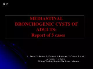
Benign Cysts • Most Common in Middle mediastinum • 20% of mediastinal masses • Less common in Korea • Usually asymptomatic • Bronchogenic cyst(32%), pericardial cyst(35%), enteric cyst(12%), thymic cyst, and thoracic duct cyst
Pericardial Cyst • Thin-walled, mesothelial cell lining • most common in Right C-P angle • Simple cysts are almost always asymptomatic • Rare cardiac impingement
Bronchogenic Cysts • 30 - 60% of all mediastinal cysts • Lined by ciliated respiratory epithelium • May contain cartilages or mucous • Communicate with tracheobronchial trees • May become infected • Wheezing, dyspnea, recurrent pulmonary infections